Skip to main content
Side panel
SPECIALTIES
Cataracts and Intraocular Lenses
Comprehensive Ophthalmology
Corneal and External Diseases
Glaucoma
Neuro-Ophthalmology
Ocular Oncology
Ophthalmic Pathology
Ophthalmic Plastic and Reconstructive Surgery
Pediatric Ophthalmology and Strabismus
Retina and Vitreous Diseases
Uveitis
FILTER BY
Online Grand Rounds
Medical Student Education
Ophthalmic Images
Other Lectures
Surgical Videos
SEARCH
More
Site-wide search
Search
Close
Perform search
Toggle search input
Log in
SPECIALTIES
Collapse
Expand
Cataracts and Intraocular Lenses
Comprehensive Ophthalmology
Corneal and External Diseases
Glaucoma
Neuro-Ophthalmology
Ocular Oncology
Ophthalmic Pathology
Ophthalmic Plastic and Reconstructive Surgery
Pediatric Ophthalmology and Strabismus
Retina and Vitreous Diseases
Uveitis
FILTER BY
Collapse
Expand
Online Grand Rounds
Medical Student Education
Ophthalmic Images
Other Lectures
Surgical Videos
SEARCH
Search
Course name
Specialty
-Any-
Cataracts and Intraocular Lenses
Comprehensive Ophthalmology
Corneal and External Diseases
Glaucoma
Neuro-Ophthalmology
Ocular Oncology
Ophthalmic Pathology
Ophthalmic Plastic and Reconstructive Surgery
Pediatric Ophthalmology and Strabismus
Retina and Vitreous Diseases
Uveitis
Other
Type
-Any-
Online Grand Rounds
Medical Student Education
Ophthalmic Images
Other Lectures
Surgical Videos
Include in Catalogue?
-Any-
No
Yes
Presenter(s)
Faculty Discussant(s)
Original Contributor(s)
Discussant(s)
Tags
Course name
|
Date
1
Page 1
2
Page 2
3
Page 3
4
Page 4
5
Page 5
6
Page 6
7
Page 7
8
Page 8
9
Page 9
10
Page 10
…
65
Page 65
»
Next page
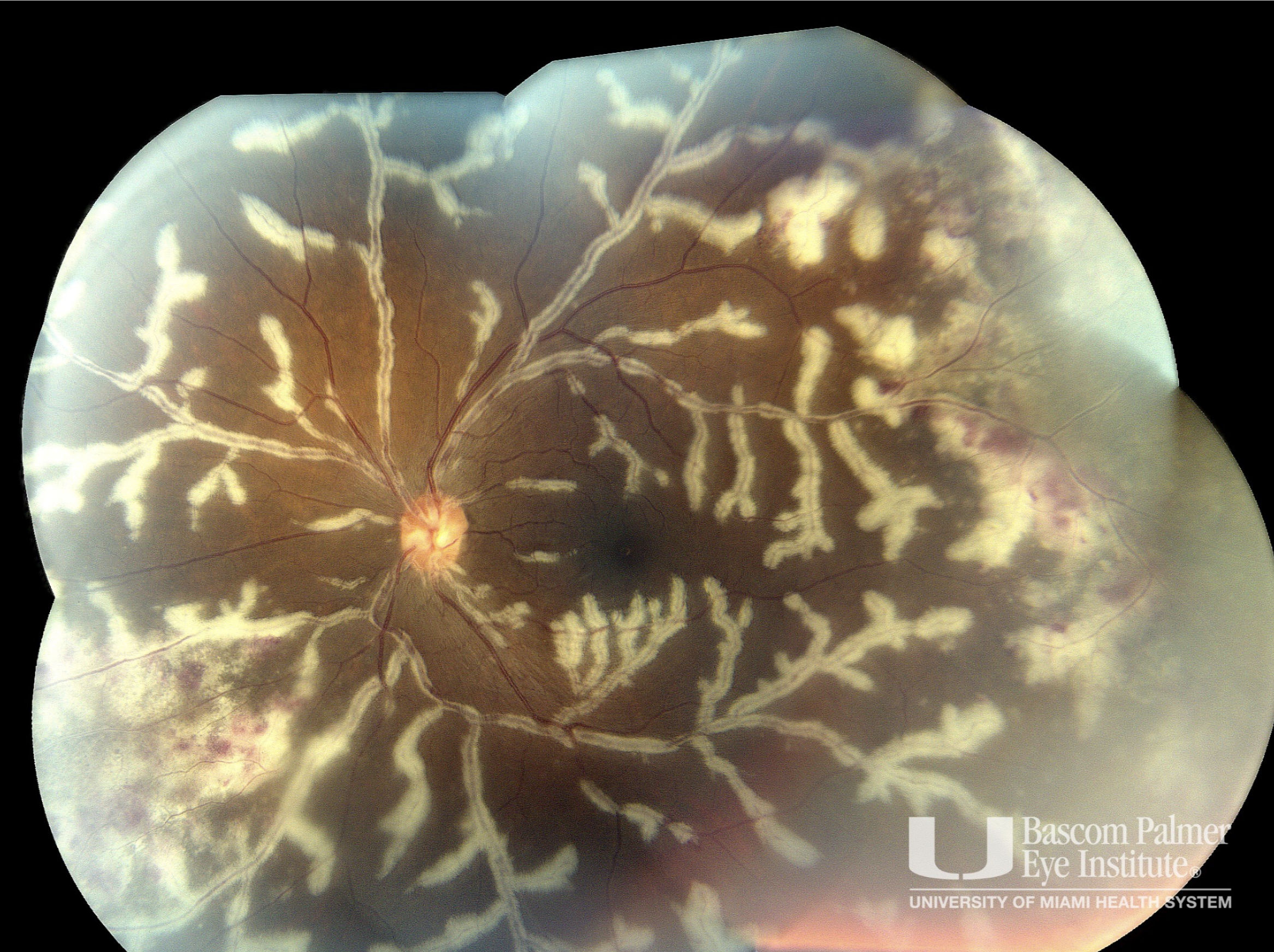
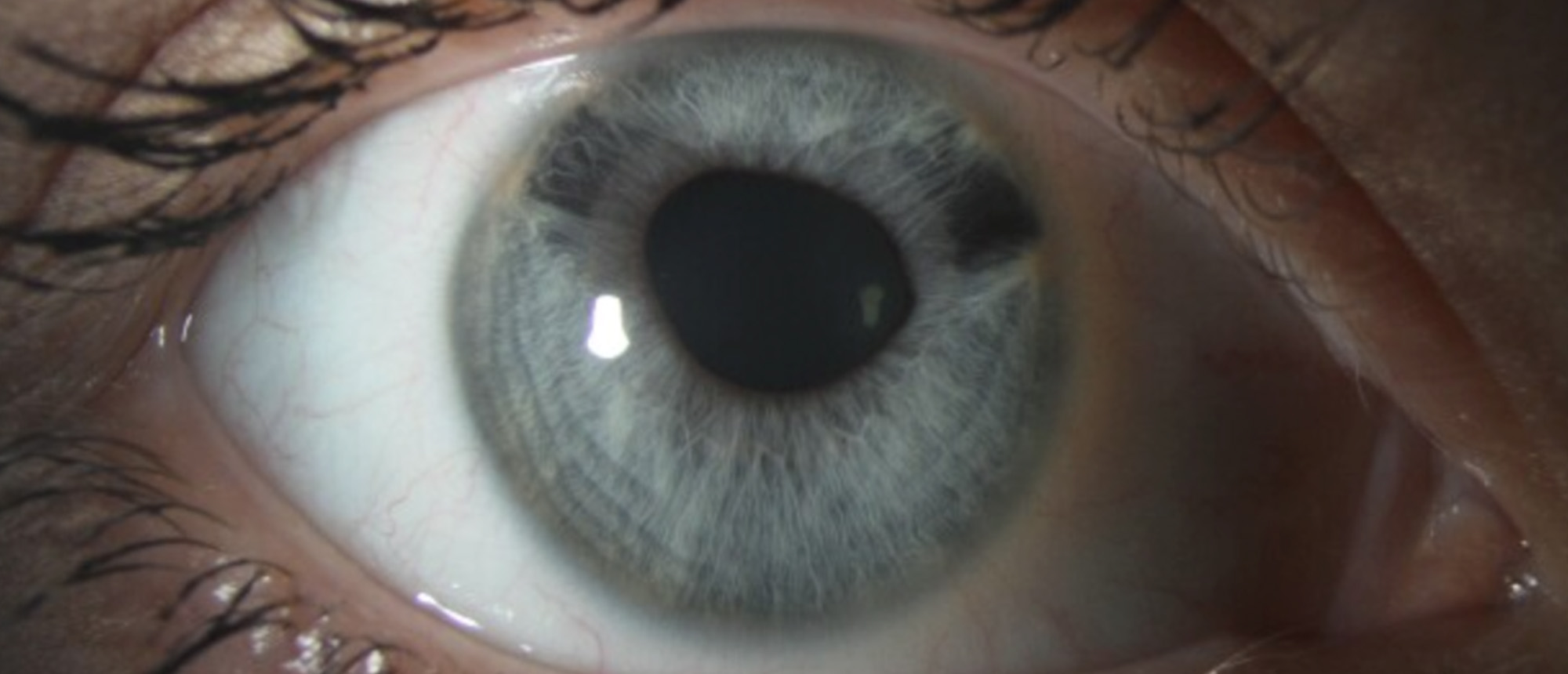
Uveitis Atlas
Specialty:
Uveitis
Type
:
Ophthalmic Images
Include in Catalogue?
:
No
Original Contributor(s)
:
Janet L. Davis, MD
The Role of Choroidal Perfusion in Age-Related Macular Degeneration
Specialty:
Retina and Vitreous Diseases
Type
:
Other Lectures
Include in Catalogue?
:
Yes
Original Contributor(s)
:
Philip J. Rosenfeld, MD, PhD
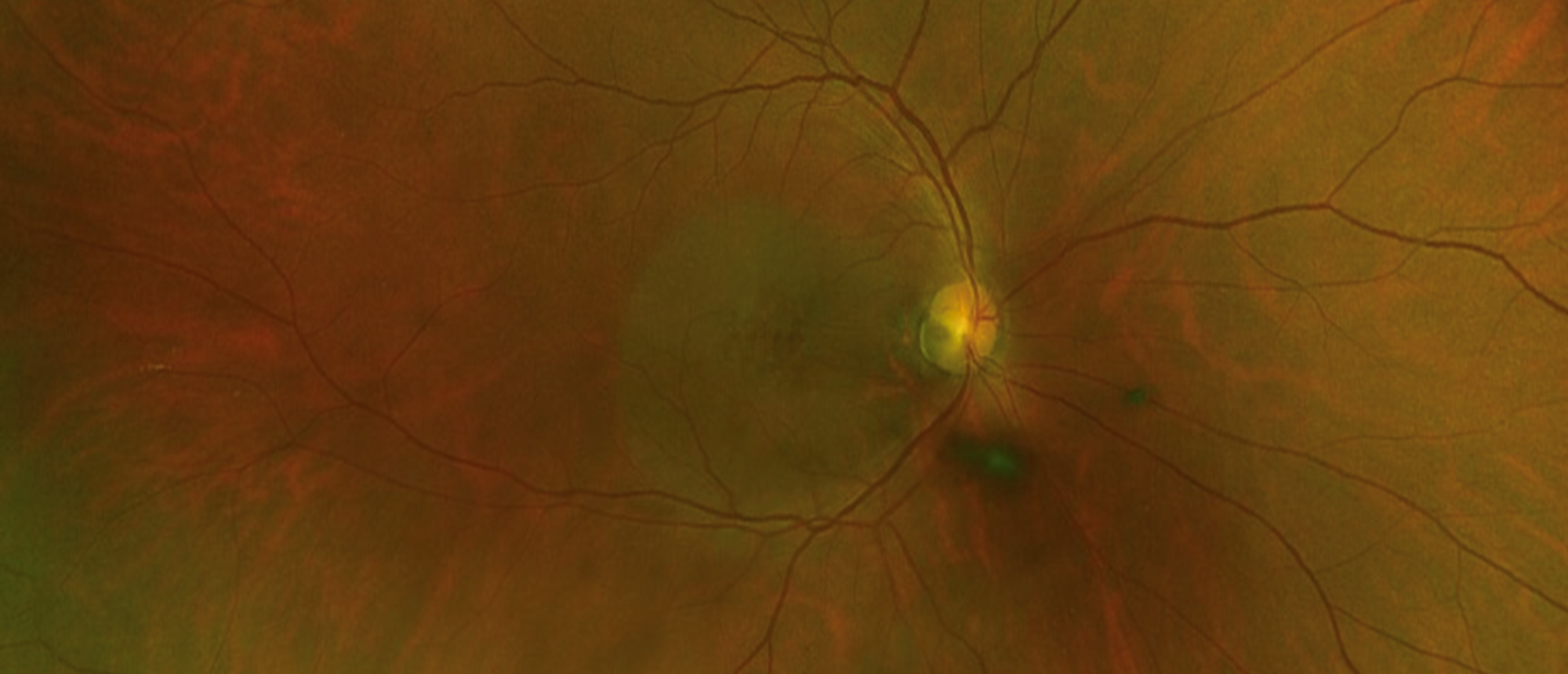
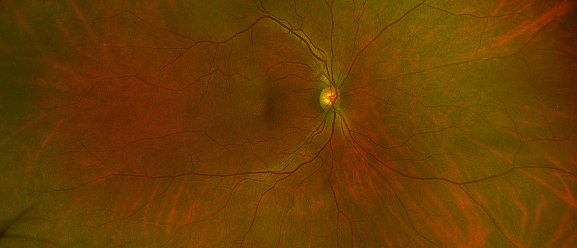
Optic Disc Pit Maculopathy
Include in Catalogue?
:
No
Presenter(s)
:
Bryant A. Menke, MD
Discussant(s)
:
Landon J. Rohowetz, MD, MA
Optic Disc Pit Maculopathy
Include in Catalogue?
:
No
Presenter(s)
:
Bryant A. Menke, MD
Discussant(s)
:
Landon J. Rohowetz, MD, MA
Optic Disc Pit Maculopathy
Specialty:
Retina and Vitreous Diseases
Type
:
Online Grand Rounds
Include in Catalogue?
:
Yes
Presenter(s)
:
Bryant A. Menke, MD
Discussant(s)
:
Landon J. Rohowetz, MD, MA
The Impact of Artificial Intelligence on Ophthalmology - Foundations, Evolution, and Clinical Reality
Type
:
Other Lectures
Include in Catalogue?
:
Yes
Original Contributor(s)
:
André Romano, MD
Langerhans Cell Histiocytosis
Include in Catalogue?
:
No
Presenter(s)
:
Mathew J. McSoley, MD, MPH
Faculty Discussant(s)
:
Sander R. Dubovy, MD; David T. Tse, MD, FACS
Langerhans Cell Histiocytosis
Include in Catalogue?
:
No
Presenter(s)
:
Mathew J. McSoley, MD, MPH
Faculty Discussant(s)
:
Sander R. Dubovy, MD; David T. Tse, MD, FACS
Langerhans Cell Histiocytosis
Specialty:
Ocular Oncology
Type
:
Online Grand Rounds
Include in Catalogue?
:
Yes
Presenter(s)
:
Mathew J. McSoley, MD, MPH
Faculty Discussant(s)
:
Sander R. Dubovy, MD; David T. Tse, MD, FACS
Endogenous Endophthalmitis and Subretinal Abscess Secondary to Klebsiella Pneumonia Bacteremia
Include in Catalogue?
:
No
Presenter(s)
:
Victor Sanchez, MD
Discussant(s)
:
Lauren C. Kiryakoza, MD
Endogenous Endophthalmitis and Subretinal Abscess Secondary to Klebsiella Pneumonia Bacteremia
Include in Catalogue?
:
No
Presenter(s)
:
Victor Sanchez, MD
Discussant(s)
:
Lauren C. Kiryakoza, MD
Endogenous Endophthalmitis and Subretinal Abscess Secondary to Klebsiella Pneumonia Bacteremia
Specialty:
Retina and Vitreous Diseases
Type
:
Online Grand Rounds
Include in Catalogue?
:
Yes
Presenter(s)
:
Victor Sanchez, MD
Discussant(s)
:
Lauren C. Kiryakoza, MD
Iridocorneal Endothelial (ICE) Syndrome, with Secondary Angle Closure Glaucoma
Include in Catalogue?
:
No
Presenter(s)
:
Georges I. Guillaume, MD
Faculty Discussant(s)
:
Elena Bitrian, MD
Iridocorneal Endothelial (ICE) Syndrome, with Secondary Angle Closure Glaucoma
Specialty:
Glaucoma
Type
:
Online Grand Rounds
Include in Catalogue?
:
Yes
Presenter(s)
:
Georges I. Guillaume, MD
Faculty Discussant(s)
:
Elena Bitrian, MD
Iridocorneal Endothelial (ICE) Syndrome, with Secondary Angle Closure Glaucoma
Include in Catalogue?
:
No
Presenter(s)
:
Georges I. Guillaume, MD
Faculty Discussant(s)
:
Elena Bitrian, MD
Traumatic Anterior Crystalline Lens Dislocation with Pupillary Block
Include in Catalogue?
:
No
Presenter(s)
:
Jacob F. Smith, MD, MS
Discussant(s)
:
Lauren C. Kiryakoza, MD; Jonathan D. Tijerina, MD, MA
Traumatic Anterior Crystalline Lens Dislocation with Pupillary Block
Include in Catalogue?
:
No
Presenter(s)
:
Jacob F. Smith, MD, MS
Discussant(s)
:
Lauren C. Kiryakoza, MD; Jonathan D. Tijerina, MD, MA
Traumatic Anterior Crystalline Lens Dislocation with Pupillary Block
Specialty:
Cataracts and Intraocular Lenses
Type
:
Online Grand Rounds
Include in Catalogue?
:
Yes
Presenter(s)
:
Jacob F. Smith, MD, MS
Discussant(s)
:
Lauren C. Kiryakoza, MD; Jonathan D. Tijerina, MD, MA
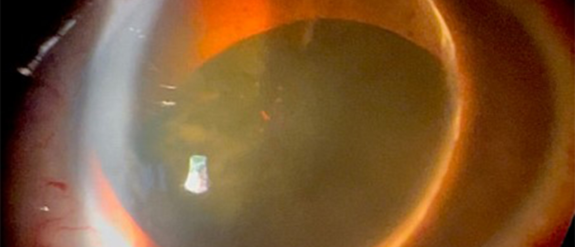
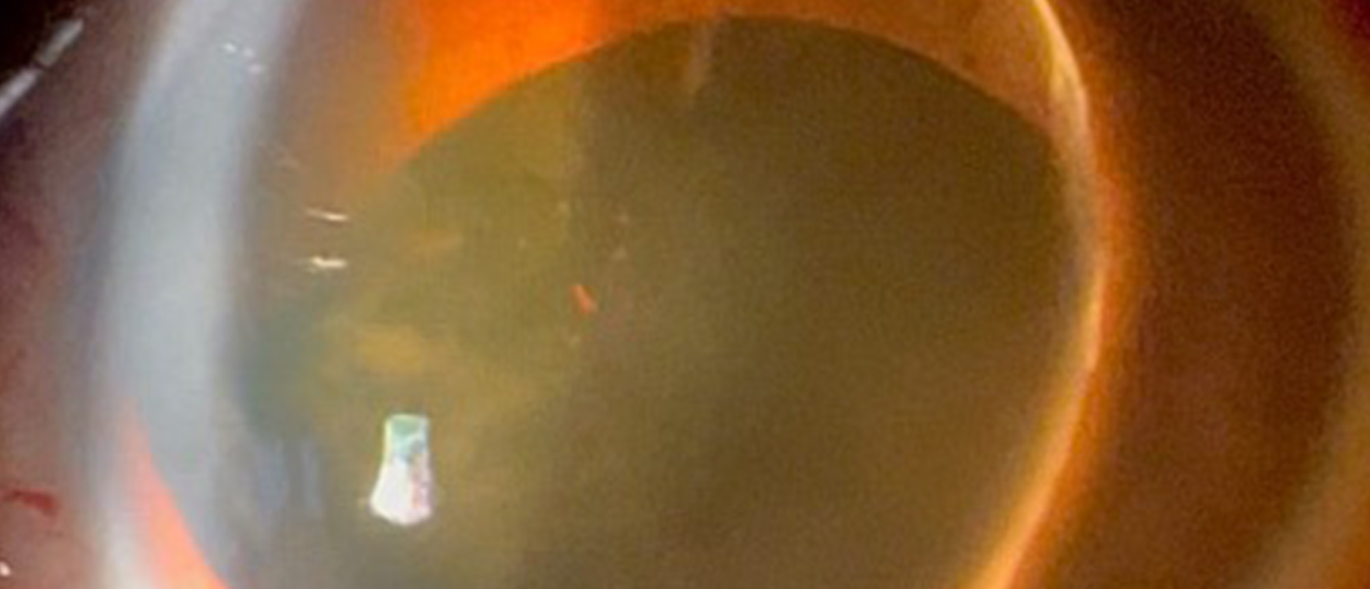
Anterior Lens Dislocation
Specialty:
Cataracts and Intraocular Lenses
Type
:
Ophthalmic Images
Include in Catalogue?
:
Yes
Original Contributor(s)
:
Lauren C. Kiryakoza, MD; Jacob F. Smith, MD, MS; Jonathan D. Tijerina, MD, MA
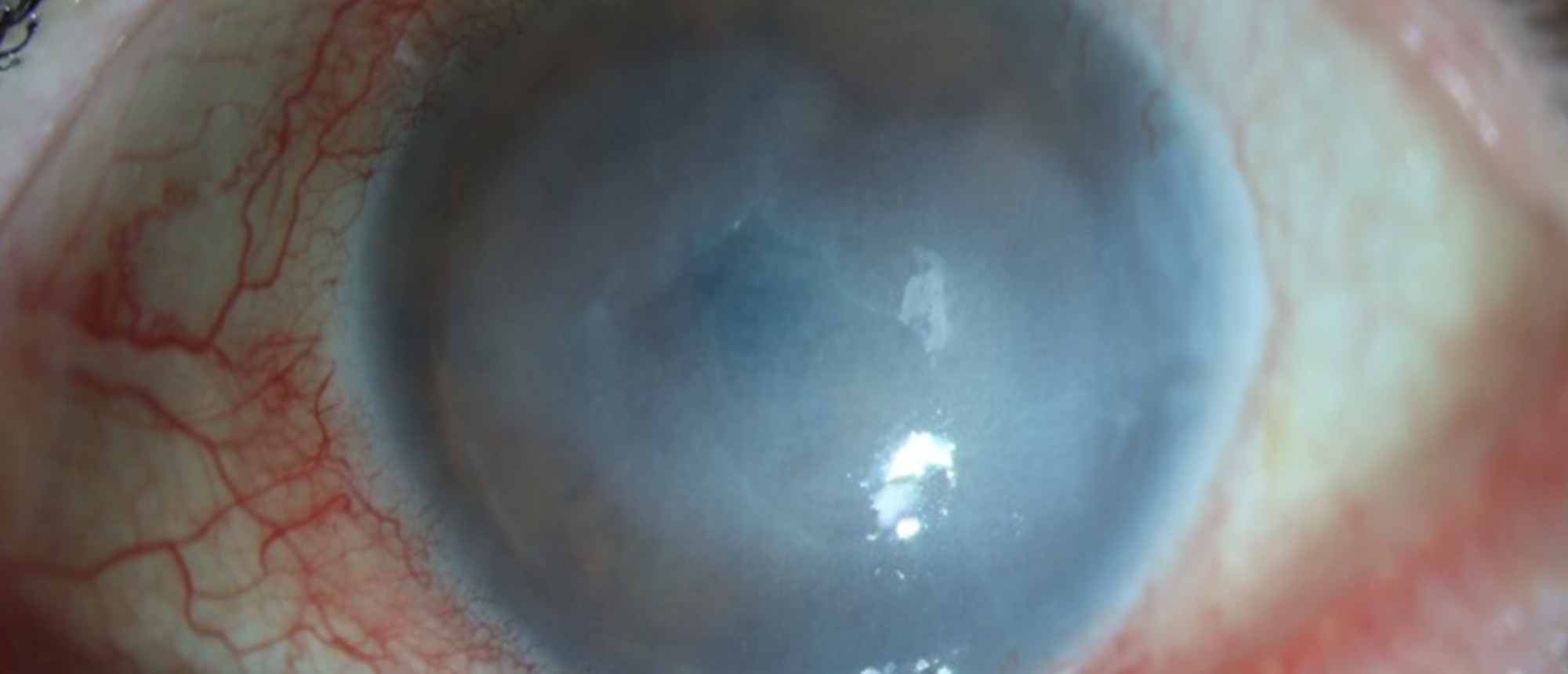
Acanthamoeba Keratitis
Include in Catalogue?
:
No
Presenter(s)
:
Helen Song, MD
Faculty Discussant(s)
:
Guillermo Amescua, MD; Darlene Miller, DHSc, MPH, CIC
1
Page 1
2
Page 2
3
Page 3
4
Page 4
5
Page 5
6
Page 6
7
Page 7
8
Page 8
9
Page 9
10
Page 10
…
65
Page 65
»
Next page
Medical Disclaimer
|
Terms of Use
|
Privacy Statement
|
HIPAA Notice of Privacy Practices
|
About Us
|
FAQs
© 2026 University of Miami Health Systems. All Rights Reserved.