
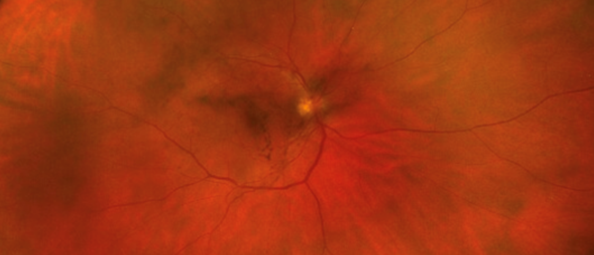
Ocular Involvement by Mycosis Fungoides / Cutaneous T Cell Lymphoma
A patient presented to the emergency department with floaters and progressive bilateral decreased vision. Examination showed bilateral vitritis with keratic precipitates, anterior chamber cell, dense vitreous haze, and snowball opacities along the inferior ora serrata. B-scan revealed dense mobile vitreous opacities without mass or retinal detachment; macular OCT was flat; fluorescein angiography showed perivascular staining with trace peripheral leakage. MRI of the brain and orbits was unremarkable. An infectious and inflammatory uveitis panel was negative, with a low angiotensin-converting enzyme and mild hypercalcemia. The picture suggested bilateral intermediate uveitis; the differential included sarcoidosis, infectious uveitides, and masquerade syndromes. Given a known systemic mature T-cell lymphoproliferative disorder, diagnostic pars plana vitrectomy was performed before immunosuppression. Undiluted and diluted vitreous were submitted for cytopathology, flow cytometry, and clonality studies. Both eyes demonstrated mature T-cell lymphoma with increased large cells; flow cytometry identified a clonal gamma-delta T-cell population, and systemic staging was negative. Treatment with intravitreal methotrexate followed by ocular radiation produced marked bilateral visual recovery. Despite excellent ocular control, a new, rapidly progressive intracranial lesion developed within weeks, and goals-of-care discussion led to a palliative approach. This case illustrates T-cell vitreoretinal lymphoma masquerading as intermediate uveitis, the primacy of tissue diagnosis, and the eye as a sentinel for central nervous system disease.
Presentation Date: 07/30/2026
Issue Date: 07/31/2026
Please log in or click on ENROLL ME to access this course.
