Skip to main content
Side panel
SPECIALTIES
Cataracts and Intraocular Lenses
Comprehensive Ophthalmology
Corneal and External Diseases
Glaucoma
Neuro-Ophthalmology
Ocular Oncology
Ophthalmic Pathology
Ophthalmic Plastic and Reconstructive Surgery
Pediatric Ophthalmology and Strabismus
Retina and Vitreous Diseases
Uveitis
FILTER BY
Online Grand Rounds
Medical Student Education
Ophthalmic Images
Other Lectures
Surgical Videos
SEARCH
More
Site-wide search
Search
Close
Perform search
Toggle search input
Log in
SPECIALTIES
Collapse
Expand
Cataracts and Intraocular Lenses
Comprehensive Ophthalmology
Corneal and External Diseases
Glaucoma
Neuro-Ophthalmology
Ocular Oncology
Ophthalmic Pathology
Ophthalmic Plastic and Reconstructive Surgery
Pediatric Ophthalmology and Strabismus
Retina and Vitreous Diseases
Uveitis
FILTER BY
Collapse
Expand
Online Grand Rounds
Medical Student Education
Ophthalmic Images
Other Lectures
Surgical Videos
SEARCH
Search
Course name
Specialty
-Any-
Cataracts and Intraocular Lenses
Comprehensive Ophthalmology
Corneal and External Diseases
Glaucoma
Neuro-Ophthalmology
Ocular Oncology
Ophthalmic Pathology
Ophthalmic Plastic and Reconstructive Surgery
Pediatric Ophthalmology and Strabismus
Retina and Vitreous Diseases
Uveitis
Other
Type
-Any-
Online Grand Rounds
Medical Student Education
Ophthalmic Images
Other Lectures
Surgical Videos
Include in Catalogue?
-Any-
No
Yes
Presenter(s)
Faculty Discussant(s)
Original Contributor(s)
Discussant(s)
Tags
Course name
|
Date
«
Previous page
1
Page 1
…
51
Page 51
52
Page 52
53
Page 53
54
Page 54
55
Page 55
56
Page 56
57
Page 57
58
Page 58
59
Page 59
60
Page 60
…
65
Page 65
»
Next page
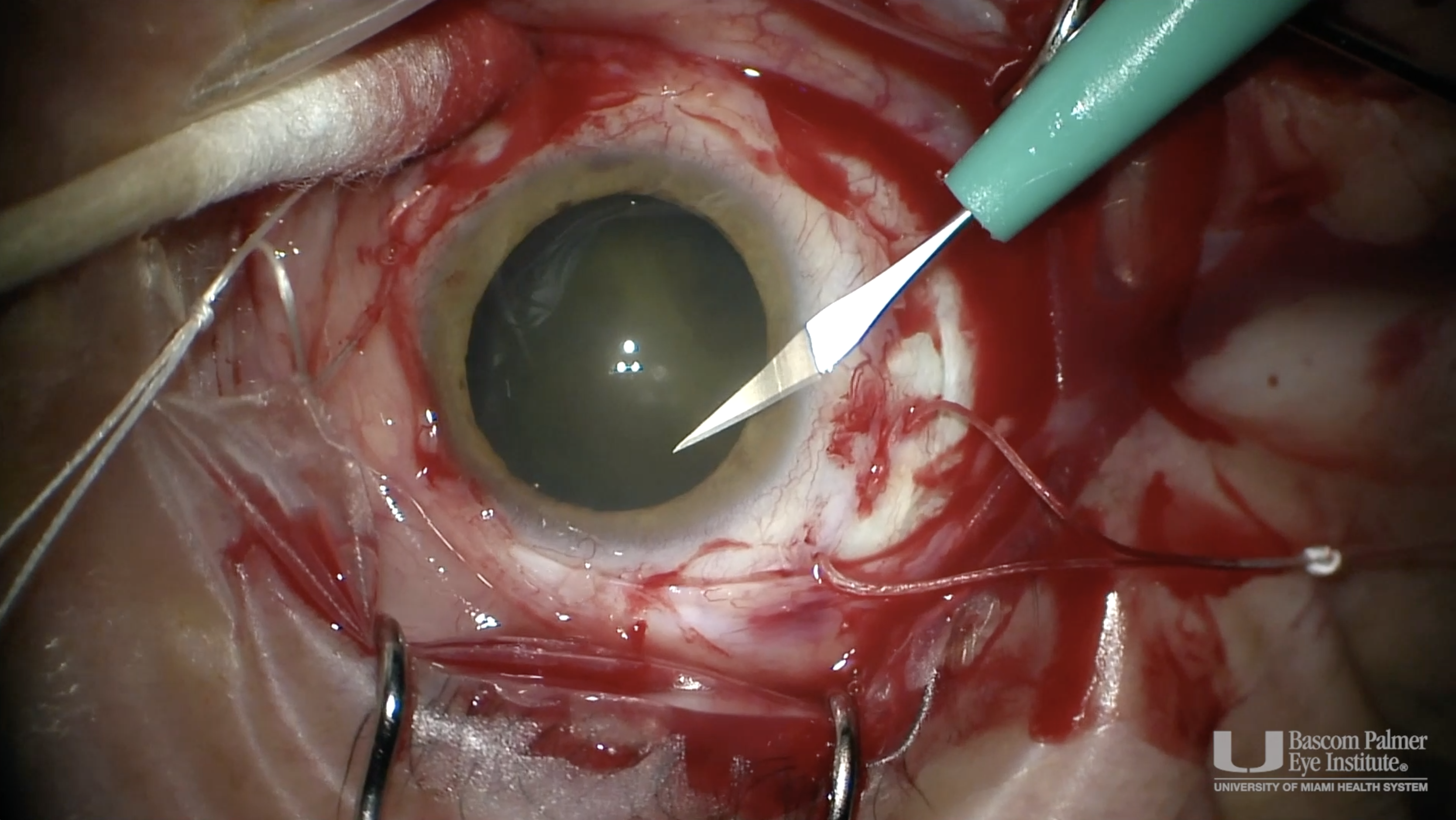
Glaucoma Drainage Device Exposure
Specialty:
Glaucoma
Type
:
Ophthalmic Images
Include in Catalogue?
:
Yes
Original Contributor(s)
:
Yasmin Islam, MD
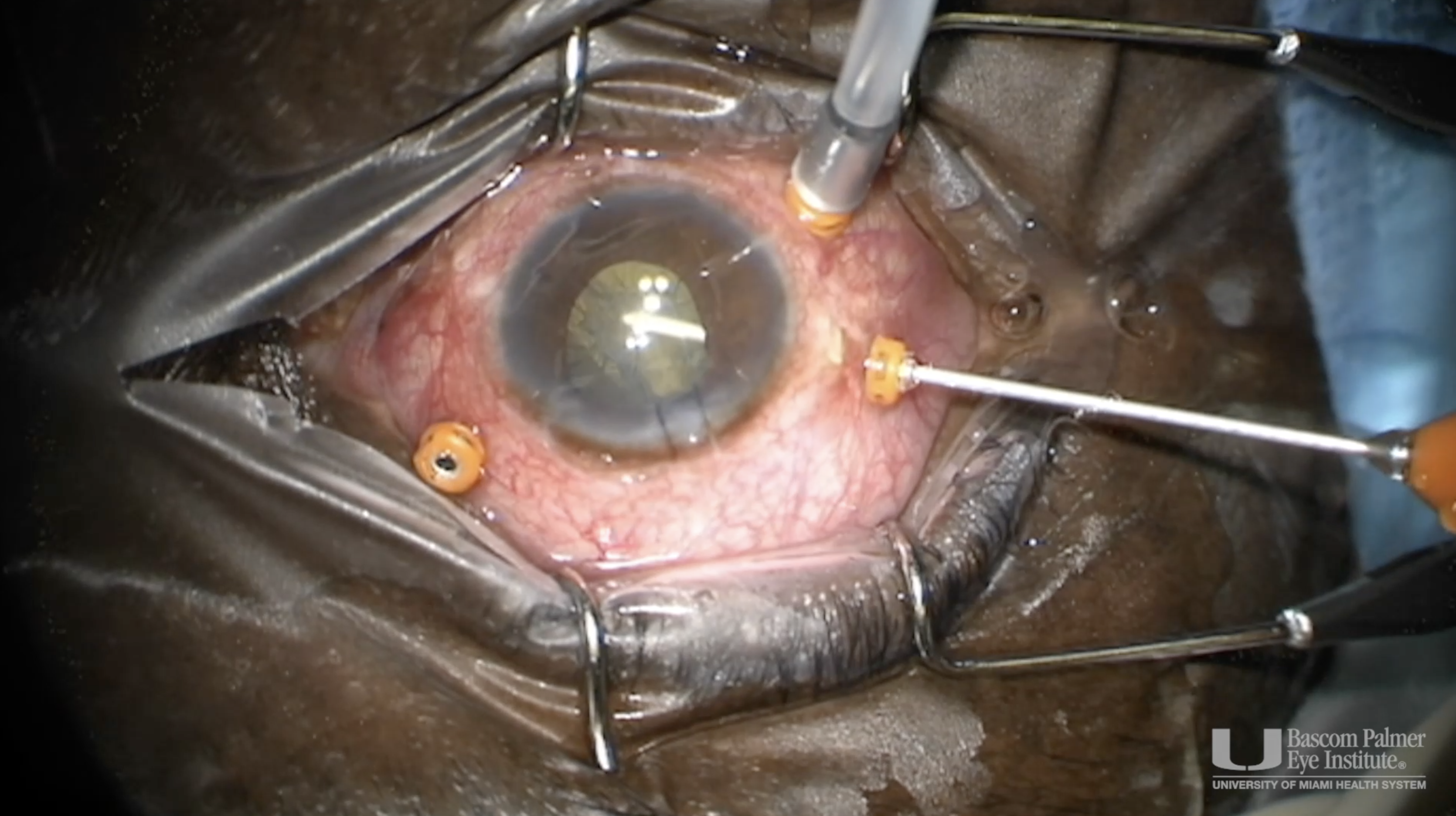
Glaucoma Drainage Device Revision
Specialty:
Glaucoma
Type
:
Surgical Videos
Include in Catalogue?
:
Yes
Original Contributor(s)
:
David S. Greenfield, MD; Yasmin Islam, MD
Saving Sight via Site-Directed Cell Trafficking
Specialty:
Other
Type
:
Other Lectures
Include in Catalogue?
:
Yes
Presenter(s)
:
Robert Sackstein, MD, PhD
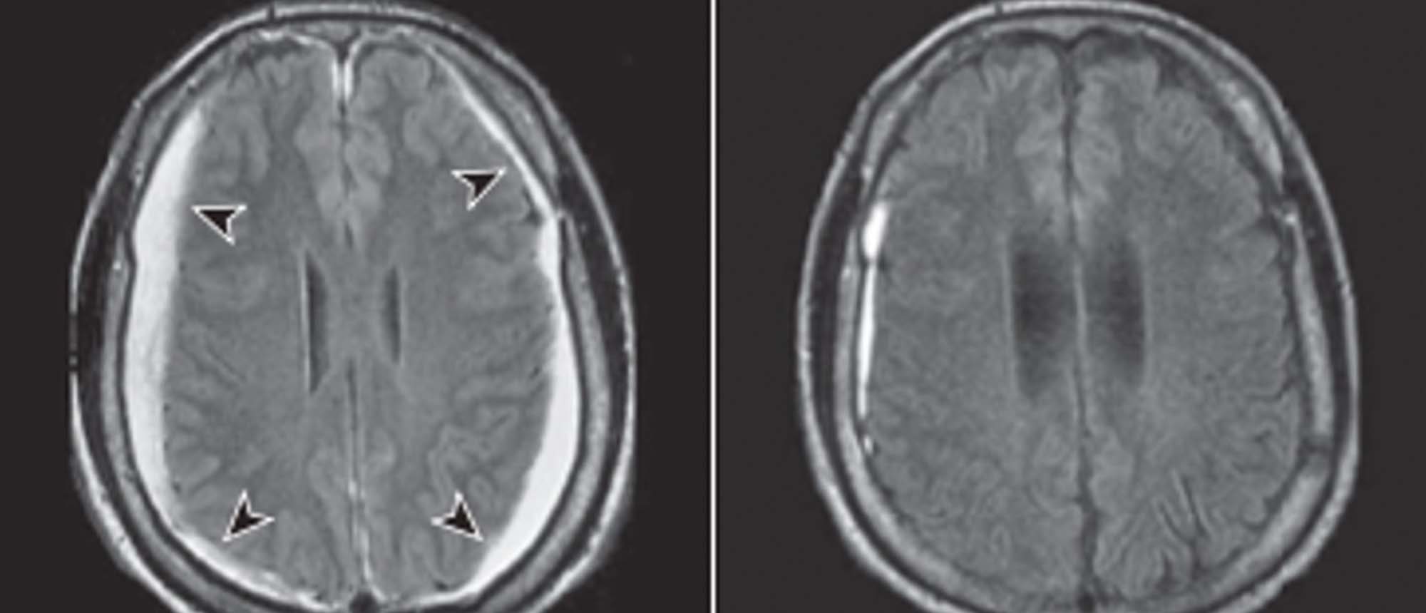
Spontaneous Intracranial Hypotension
Include in Catalogue?
:
No
Presenter(s)
:
Mariam S. Vila-Delgado, MD
Faculty Discussant(s)
:
Byron L. Lam, MD
Spontaneous Intracranial Hypotension
Specialty:
Neuro-Ophthalmology
Type
:
Online Grand Rounds
Include in Catalogue?
:
Yes
Presenter(s)
:
Mariam S. Vila-Delgado, MD
Faculty Discussant(s)
:
Byron L. Lam, MD
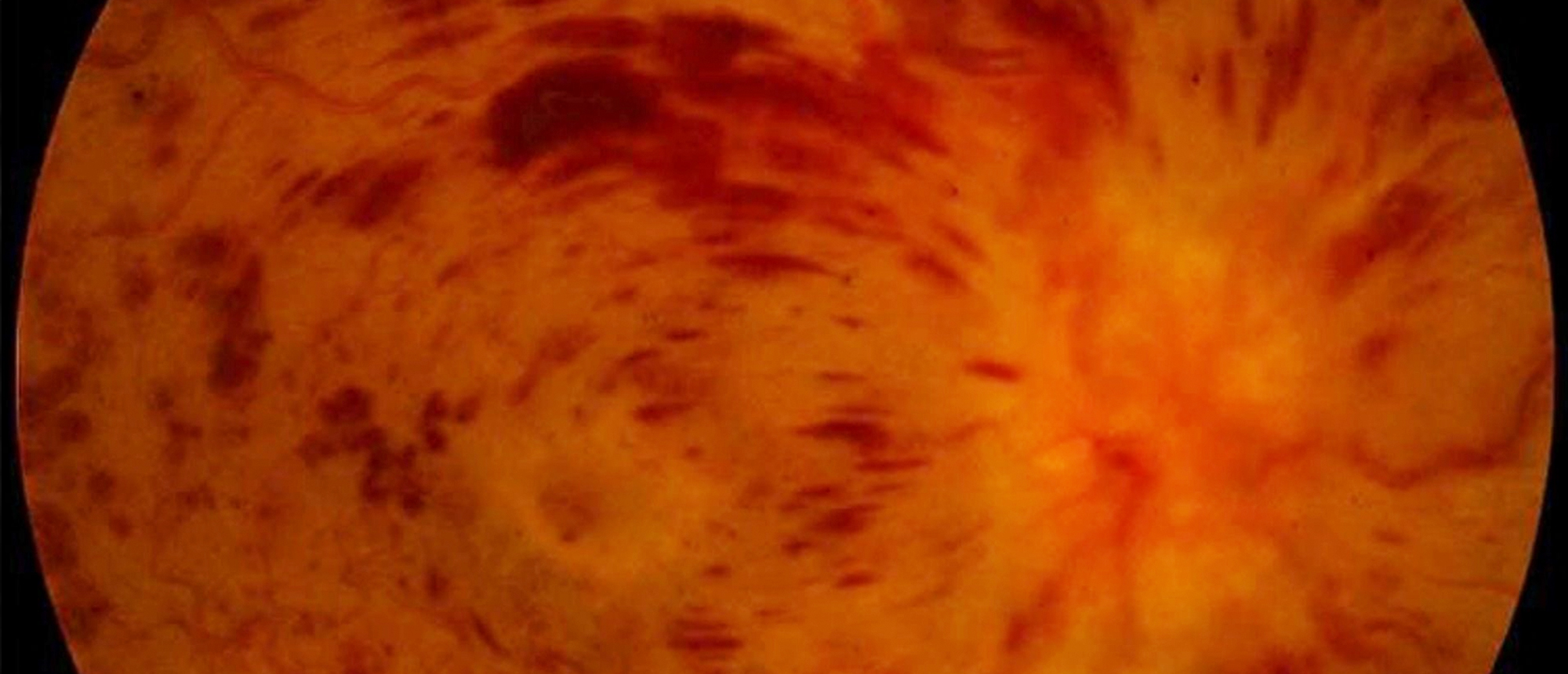
Central Retinal Vein Occlusion in Patient with MTHFR Mutation
Specialty:
Retina and Vitreous Diseases
Type
:
Online Grand Rounds
Include in Catalogue?
:
Yes
Presenter(s)
:
Julia L. Hudson, MD
Faculty Discussant(s)
:
Audina M. Berrocal, MD
Familial Exudative Vitreoretinopathy
Specialty:
Pediatric Ophthalmology and Strabismus
Type
:
Online Grand Rounds
Include in Catalogue?
:
Yes
Presenter(s)
:
Michael J. Venincasa, MD
Faculty Discussant(s)
:
Audina M. Berrocal, MD
Finances and Investing for Young, Mid-Career, and Late-Career Physicians
Specialty:
Other
Type
:
Other Lectures
Include in Catalogue?
:
Yes
Presenter(s)
:
Myron Tanenbaum, MD
Addressing Health Inequities and Social Determinants of Health: an Ophthalmologist's Perspective
Specialty:
Other
Type
:
Other Lectures
Include in Catalogue?
:
Yes
Presenter(s)
:
Fasika A. Woreta, MD, MPH
Closing the Health Gap: The Role of Diversity in Ophthalmology
Specialty:
Other
Type
:
Other Lectures
Include in Catalogue?
:
Yes
Presenter(s)
:
Basil K. Williams, Jr., MD
Queratitis por Hongos
Specialty:
Corneal and External Diseases
Type
:
Other Lectures
Include in Catalogue?
:
Yes
Presenter(s)
:
Eduardo Alfonso, MD
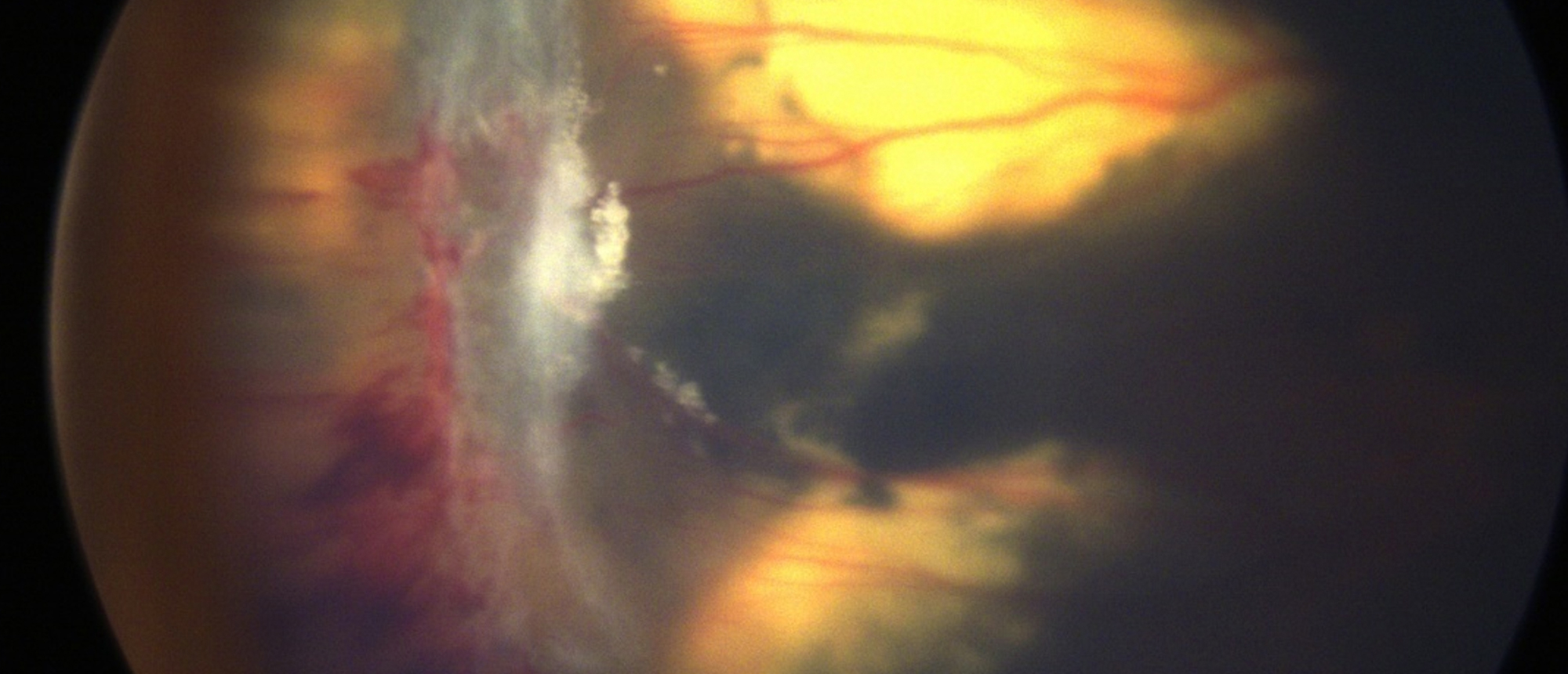
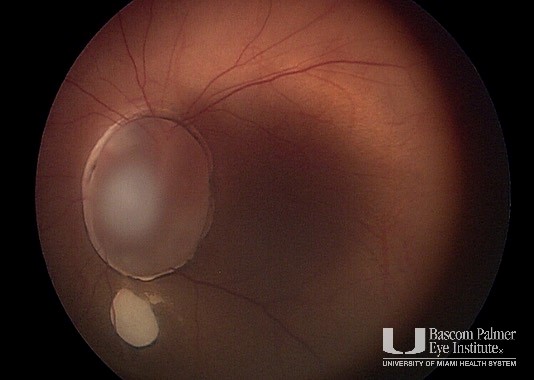
Optic Nerve Cyst and Coloboma
Specialty:
Pediatric Ophthalmology and Strabismus
Type
:
Ophthalmic Images
Include in Catalogue?
:
Yes
Original Contributor(s)
:
Audina M. Berrocal, MD; Nimesh A. Patel, MD
57th Annual Residents' Day
Specialty:
Other
Type
:
Other Lectures
Include in Catalogue?
:
Yes
Original Contributor(s)
:
Harry W. Flynn, Jr., MD; Steven J. Gedde, MD
Epithelial Downgrowth
Specialty:
Cataracts and Intraocular Lenses
Type
:
Online Grand Rounds
Include in Catalogue?
:
Yes
Presenter(s)
:
Ghada J. AlBayyat, MD
Faculty Discussant(s)
:
Sander R. Dubovy, MD; Carol L. Karp, MD
External Drainage of Choroidal Melanoma Retinal Detachment
Specialty:
Retina and Vitreous Diseases
Type
:
Surgical Videos
Include in Catalogue?
:
Yes
Original Contributor(s)
:
J. Daniel Diaz, MD; J. William Harbour, MD; Nimesh A. Patel, MD
Sub-Retina Band Removal in PVR Detachment
Specialty:
Retina and Vitreous Diseases
Type
:
Surgical Videos
Include in Catalogue?
:
Yes
Original Contributor(s)
:
Nimesh A. Patel, MD; Jayanth Sridhar, MD
Orbital Cysts
Specialty:
Ophthalmic Plastic and Reconstructive Surgery
Type
:
Ophthalmic Images
Include in Catalogue?
:
Yes
Original Contributor(s)
:
Shanlee M. Stevens, MD
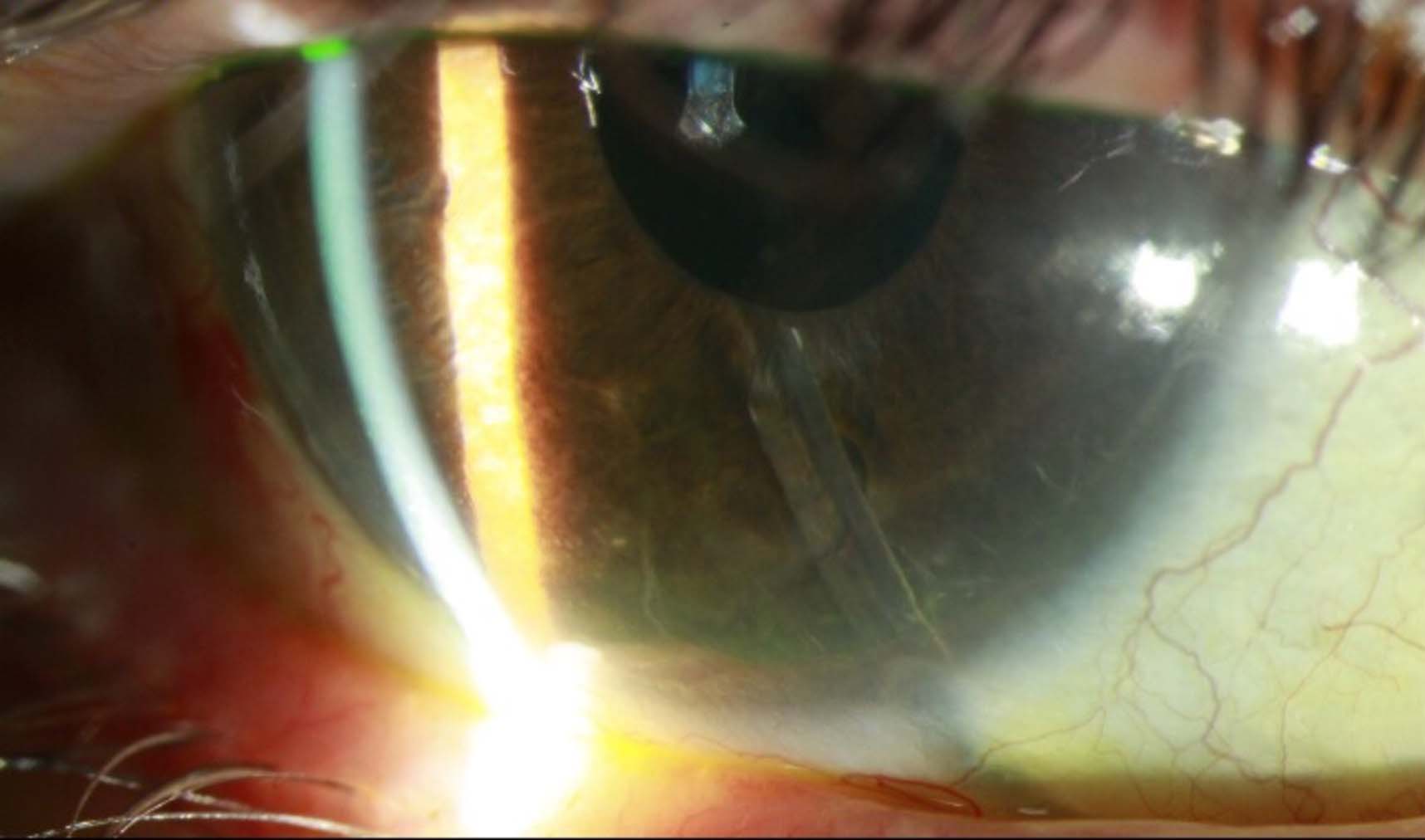
Phacolytic Glaucoma
Specialty:
Cataracts and Intraocular Lenses
Type
:
Ophthalmic Images
Include in Catalogue?
:
Yes
Original Contributor(s)
:
Sander R. Dubovy, MD; Thomas A. Lazzarini, MD; Nathan L. Scott, MD, MPP; Justin H. Townsend, MD
Reflections on the COVID-19 Pandemic and its Impact on Ophthalmology
Specialty:
Other
Type
:
Other Lectures
Include in Catalogue?
:
Yes
Presenter(s)
:
Paul Sternberg, Jr., MD
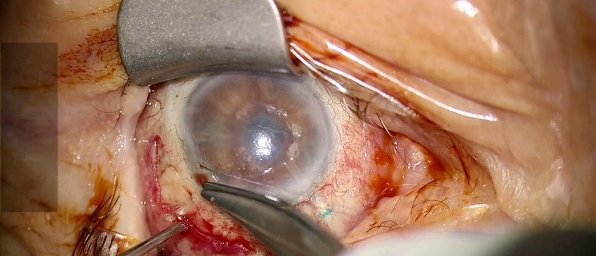
Toxoplasmosis Biopsy
Specialty:
Retina and Vitreous Diseases
Type
:
Surgical Videos
Include in Catalogue?
:
Yes
Original Contributor(s)
:
Joshua Uhr, MD; Nathan L. Scott, MD, MPP
«
Previous page
1
Page 1
…
51
Page 51
52
Page 52
53
Page 53
54
Page 54
55
Page 55
56
Page 56
57
Page 57
58
Page 58
59
Page 59
60
Page 60
…
65
Page 65
»
Next page
Medical Disclaimer
|
Terms of Use
|
Privacy Statement
|
HIPAA Notice of Privacy Practices
|
About Us
|
FAQs
© 2026 University of Miami Health Systems. All Rights Reserved.