Bascom Palmer
  • Calendar
  • SPECIALTIES
    Cataracts and Intraocular Lenses Comprehensive Ophthalmology Corneal and External Diseases Glaucoma Neuro-Ophthalmology Ocular Oncology Ophthalmic Pathology Ophthalmic Plastic and Reconstructive Surgery Pediatric Ophthalmology and Strabismus Retina and Vitreous Diseases Uveitis
  • FILTER BY
    Online Grand Rounds Medical Student Education Ophthalmic Images Other Lectures Surgical Videos
  • SEARCH
  • More
Site-wide search Close
Toggle search input
guest access
Log in
Bascom Palmer
Calendar SPECIALTIES Collapse Expand
Cataracts and Intraocular Lenses Comprehensive Ophthalmology Corneal and External Diseases Glaucoma Neuro-Ophthalmology Ocular Oncology Ophthalmic Pathology Ophthalmic Plastic and Reconstructive Surgery Pediatric Ophthalmology and Strabismus Retina and Vitreous Diseases Uveitis
FILTER BY Collapse Expand
Online Grand Rounds Medical Student Education Ophthalmic Images Other Lectures Surgical Videos
SEARCH
Expand all Collapse all
  1. Dashboard
  2. CN110622OI
  3. Ophthalmic Image(s)
  4. Corneal Neurotization

Corneal Neurotization

Completion requirements

Description

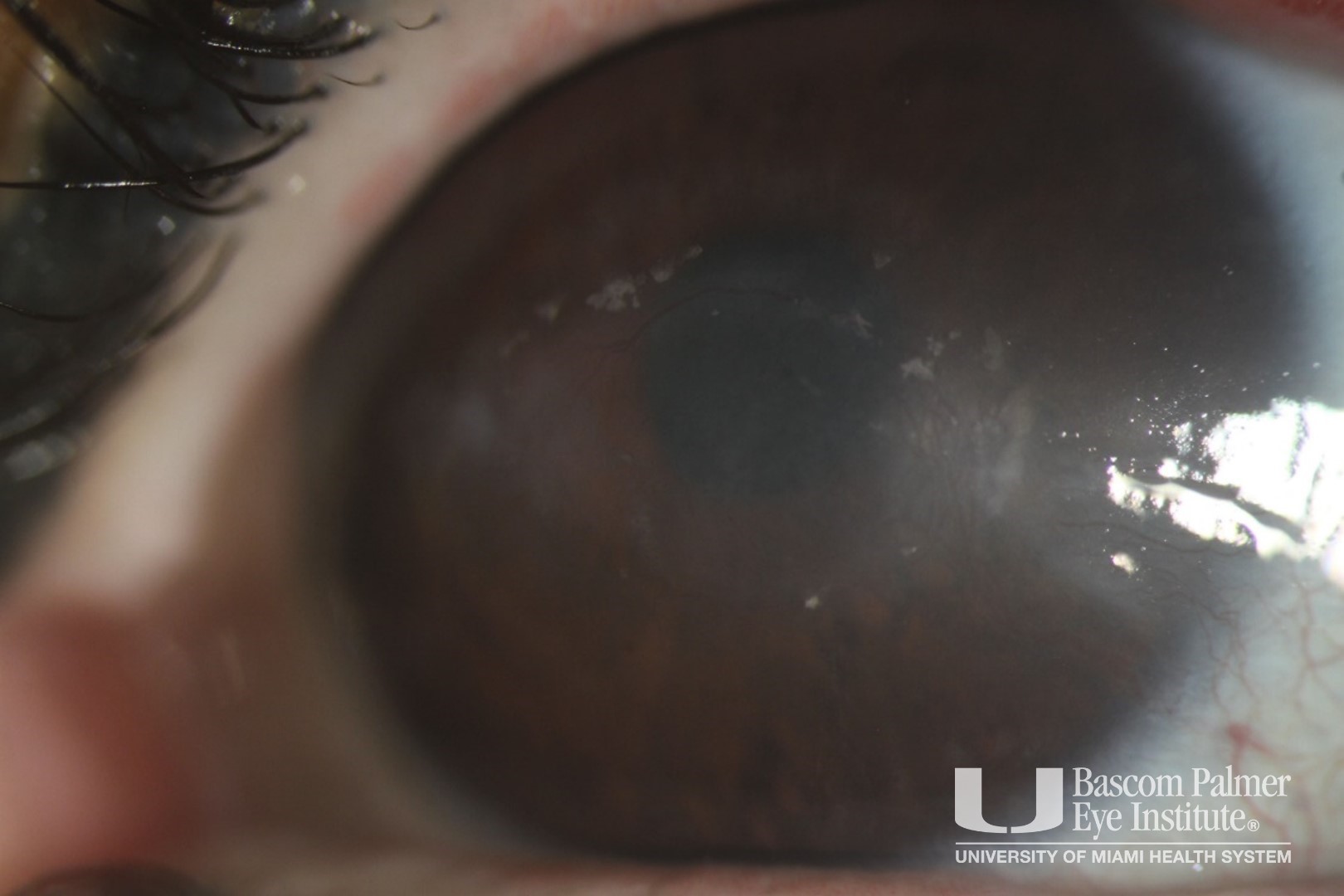
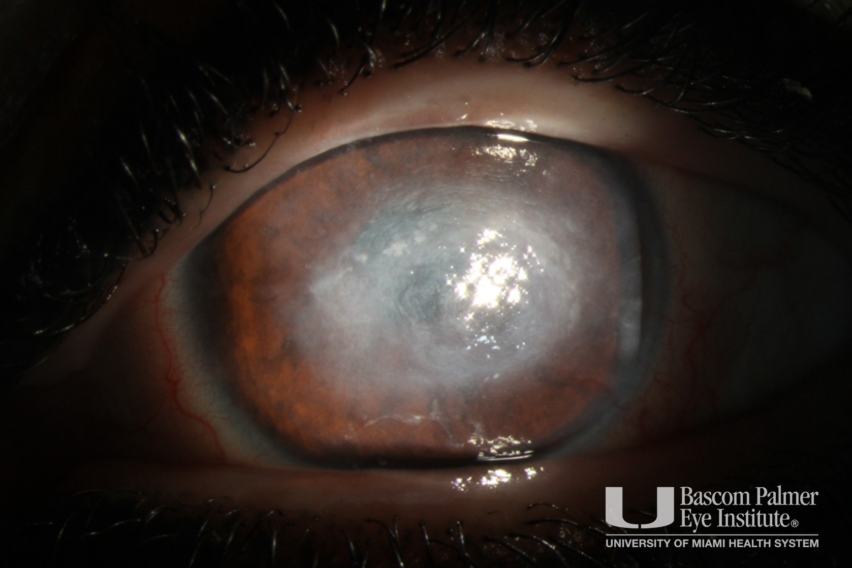
This patient presented with left-sided CN 5, 6, 7, and 8 palsies leading to multiple emergency visits for corneal erosions and infectious corneal ulcers. Prior treatment was with amniotic membrane discs, superficial keratectomy with EDTA, botox tarsorrhaphy, and a surgical ertractor reinsertion with lateral canthal tendon plication.

The patient then underwent a Corneal Neurotization procedure using an allograft to the contralateral supraorbital nerve. This was performed by Dr. Rong and Dr. Amescua. The surgeons exposes the superior orbital rim through an upper eyelid incision. They identify and dissect the supraorbital nerve, which will be transected. The contralateral upper eyelid incision is then made. The processed nerve allograft is attached to a nerve connector. The supraorbital nerve is fed into the opposite end, forming an end-to-end connection. The graft is then tunneled to the affected side. It is brought to the affected cornea, and the fascicles are inserted into the conjunctiva.

The vision improved from CF to 20/70 and the corneal epithelial defect healed.

Uploaded on: 11/06/2022


Corneal Neurotization


Corneal Neurotization


◄ Contributor: Andrew J. Rong, MD
guest access (Log in)
Data retention summary
Powered by Moodle
 
  • Medical Disclaimer
  • |
  • Terms of Use
  • |
  • Privacy Statement
  • |
  • HIPAA Notice of Privacy Practices
  • |
  • About Us
  • |
  • FAQs

© 2026 University of Miami Health Systems. All Rights Reserved.