Sequential Ultrawide Field Fundus Photo, Left Eye
Completion requirements
Description
Description
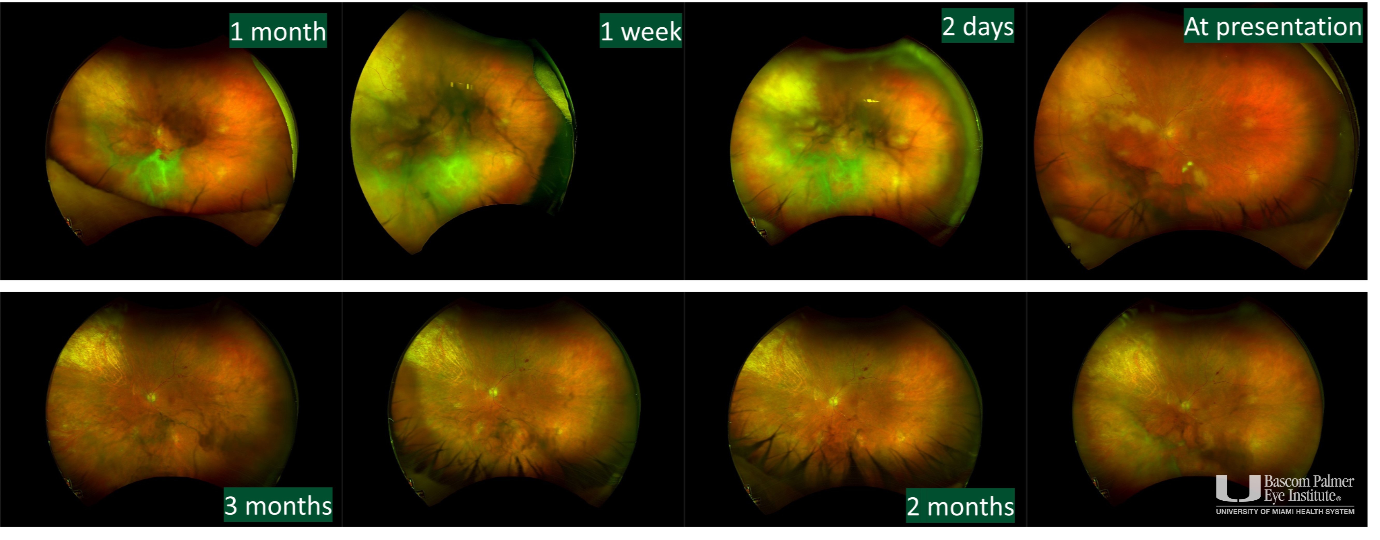
A patient with atypical presentation of toxoplasma chorioretinitis. Ultra wide field color fundus photograph showing a peripheral retinal whitening with patchy areas of active inflammation underlying a grade 2 vitreous haze in the left eye at presentation. The patient was initially suspected of acute retinal necrosis which worsened on antiviral therapy. Antibacterial monotherapy for toxoplasma retinitis was started following a confirmatory diagnosis made by polymerase chain reaction of anterior chamber fluid. Sequential photographs show significant improvement after treatment.
Uploaded on: 10/20/2023