Skip to main content
Side panel
SPECIALTIES
Cataracts and Intraocular Lenses
Comprehensive Ophthalmology
Corneal and External Diseases
Glaucoma
Neuro-Ophthalmology
Ocular Oncology
Ophthalmic Pathology
Ophthalmic Plastic and Reconstructive Surgery
Pediatric Ophthalmology and Strabismus
Retina and Vitreous Diseases
Uveitis
FILTER BY
Online Grand Rounds
Medical Student Education
Ophthalmic Images
Other Lectures
Surgical Videos
SEARCH
More
Site-wide search
Search
Close
Perform search
Toggle search input
Log in
SPECIALTIES
Collapse
Expand
Cataracts and Intraocular Lenses
Comprehensive Ophthalmology
Corneal and External Diseases
Glaucoma
Neuro-Ophthalmology
Ocular Oncology
Ophthalmic Pathology
Ophthalmic Plastic and Reconstructive Surgery
Pediatric Ophthalmology and Strabismus
Retina and Vitreous Diseases
Uveitis
FILTER BY
Collapse
Expand
Online Grand Rounds
Medical Student Education
Ophthalmic Images
Other Lectures
Surgical Videos
SEARCH
Search
Course name
Specialty
-Any-
Cataracts and Intraocular Lenses
Comprehensive Ophthalmology
Corneal and External Diseases
Glaucoma
Neuro-Ophthalmology
Ocular Oncology
Ophthalmic Pathology
Ophthalmic Plastic and Reconstructive Surgery
Pediatric Ophthalmology and Strabismus
Retina and Vitreous Diseases
Uveitis
Other
Type
-Any-
Online Grand Rounds
Medical Student Education
Ophthalmic Images
Other Lectures
Surgical Videos
Include in Catalogue?
-Any-
No
Yes
Presenter(s)
Faculty Discussant(s)
Original Contributor(s)
Discussant(s)
Tags
Course name
|
Date
«
Previous page
1
Page 1
…
49
Page 49
50
Page 50
51
Page 51
52
Page 52
53
Page 53
54
Page 54
55
Page 55
56
Page 56
57
Page 57
58
Page 58
…
65
Page 65
»
Next page
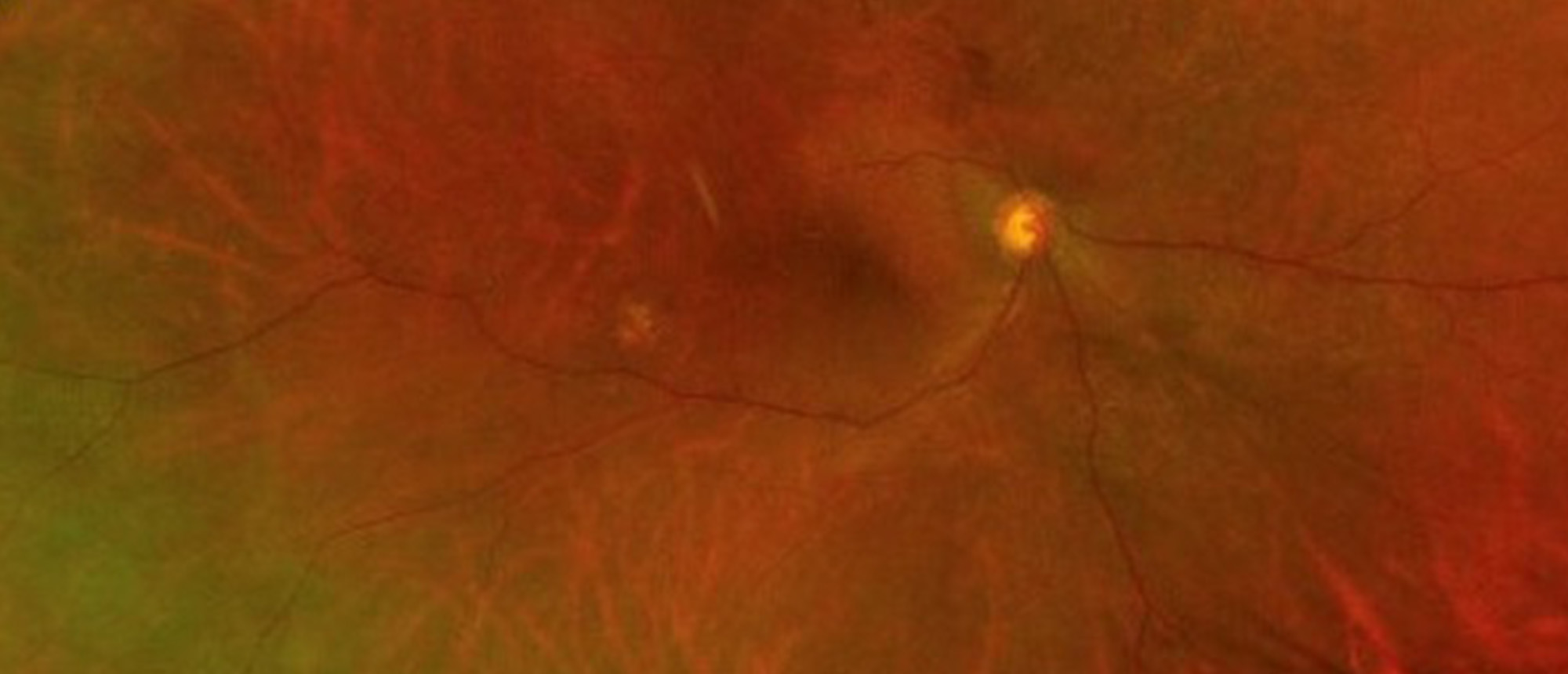
Vitreous Hemorrhage
Include in Catalogue?
:
No
Presenter(s)
:
Anne L. Kunkler, MD
Faculty Discussant(s)
:
Harry W. Flynn, Jr., MD
Keratoacanthoma
Specialty:
Corneal and External Diseases
Type
:
Ophthalmic Images
Include in Catalogue?
:
Yes
Original Contributor(s)
:
Humberto Salazar, III, MD
Leukemic Optic Nerve Infiltrate
Specialty:
Neuro-Ophthalmology
Type
:
Ophthalmic Images
Include in Catalogue?
:
Yes
Original Contributor(s)
:
Byron L. Lam, MD; Caroline L. Lieux, MD
Leukemic Optic Neuropathy
Include in Catalogue?
:
No
Presenter(s)
:
Caroline L. Lieux, MD
Faculty Discussant(s)
:
Byron L. Lam, MD
Leukemic Optic Neuropathy
Include in Catalogue?
:
No
Presenter(s)
:
Caroline L. Lieux, MD
Faculty Discussant(s)
:
Byron L. Lam, MD
Leukemic Optic Neuropathy
Specialty:
Neuro-Ophthalmology
Type
:
Online Grand Rounds
Include in Catalogue?
:
Yes
Presenter(s)
:
Caroline L. Lieux, MD
Faculty Discussant(s)
:
Byron L. Lam, MD
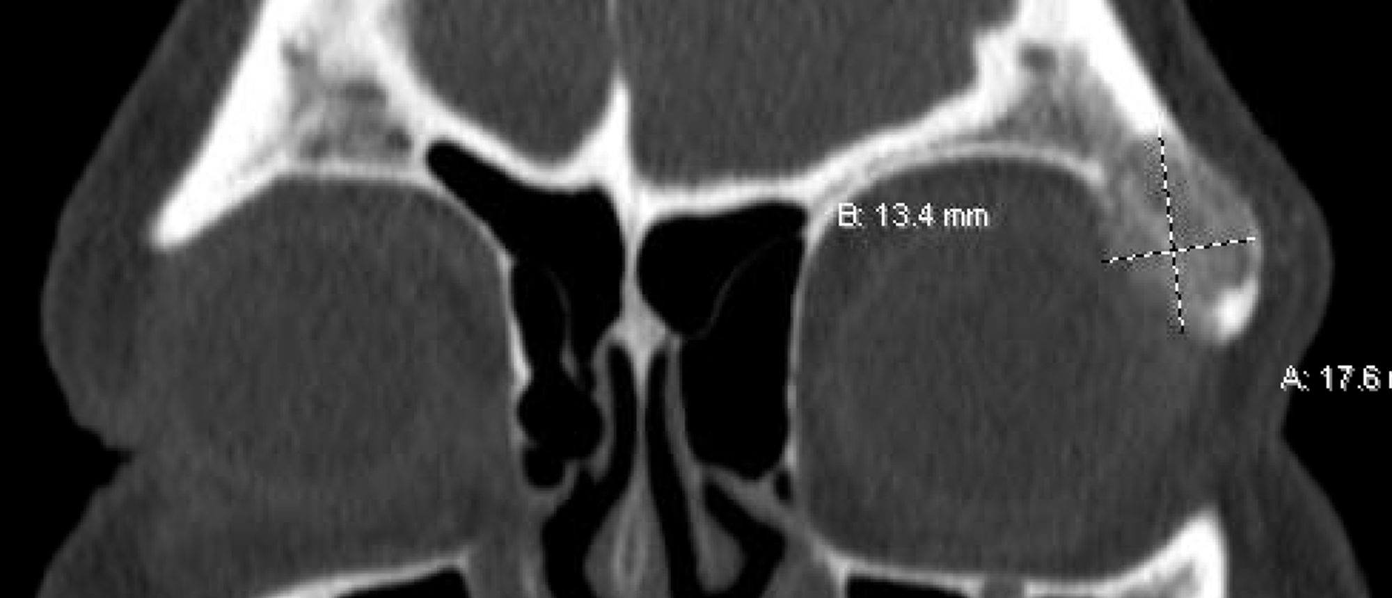
Intraosseous Hemangioma
Specialty:
Ophthalmic Plastic and Reconstructive Surgery
Type
:
Online Grand Rounds
Include in Catalogue?
:
Yes
Presenter(s)
:
Michelle M. Maeng, MD
Faculty Discussant(s)
:
Sander R. Dubovy, MD; David T. Tse, MD
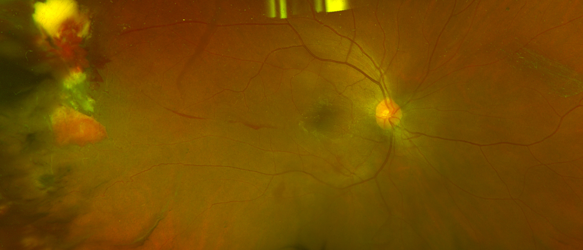
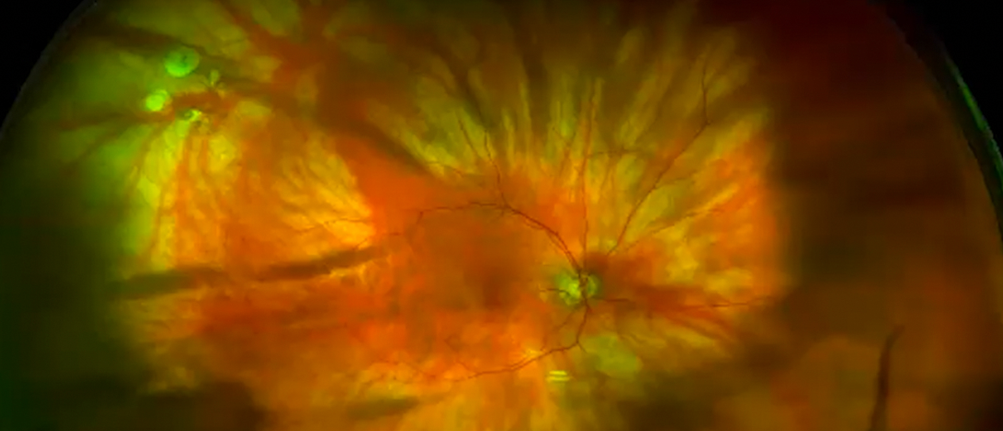
Autosomal Dominant Neovascular Inflammatory Vitreoretinopathy
Specialty:
Retina and Vitreous Diseases
Type
:
Online Grand Rounds
Include in Catalogue?
:
Yes
Presenter(s)
:
Julia L. Hudson, MD
Faculty Discussant(s)
:
Audina M. Berrocal, MD
Hydrolyzed MIRAgel Scleral Buckle
Specialty:
Ophthalmic Plastic and Reconstructive Surgery
Type
:
Online Grand Rounds
Include in Catalogue?
:
Yes
Presenter(s)
:
Anne L. Kunkler, MD
Faculty Discussant(s)
:
Audina M. Berrocal, MD; Brian C. Tse, MD
Best of Curso 2021
Specialty:
Other
Type
:
Other Lectures
Include in Catalogue?
:
Yes
Presenter(s)
:
Eduardo Alfonso, MD; Carol L. Karp, MD; Paul F. Palmberg, MD, PhD
Advances in Global Ophthalmic Education
Specialty:
Other
Type
:
Other Lectures
Include in Catalogue?
:
Yes
Presenter(s)
:
Juan F. Batlle, MD
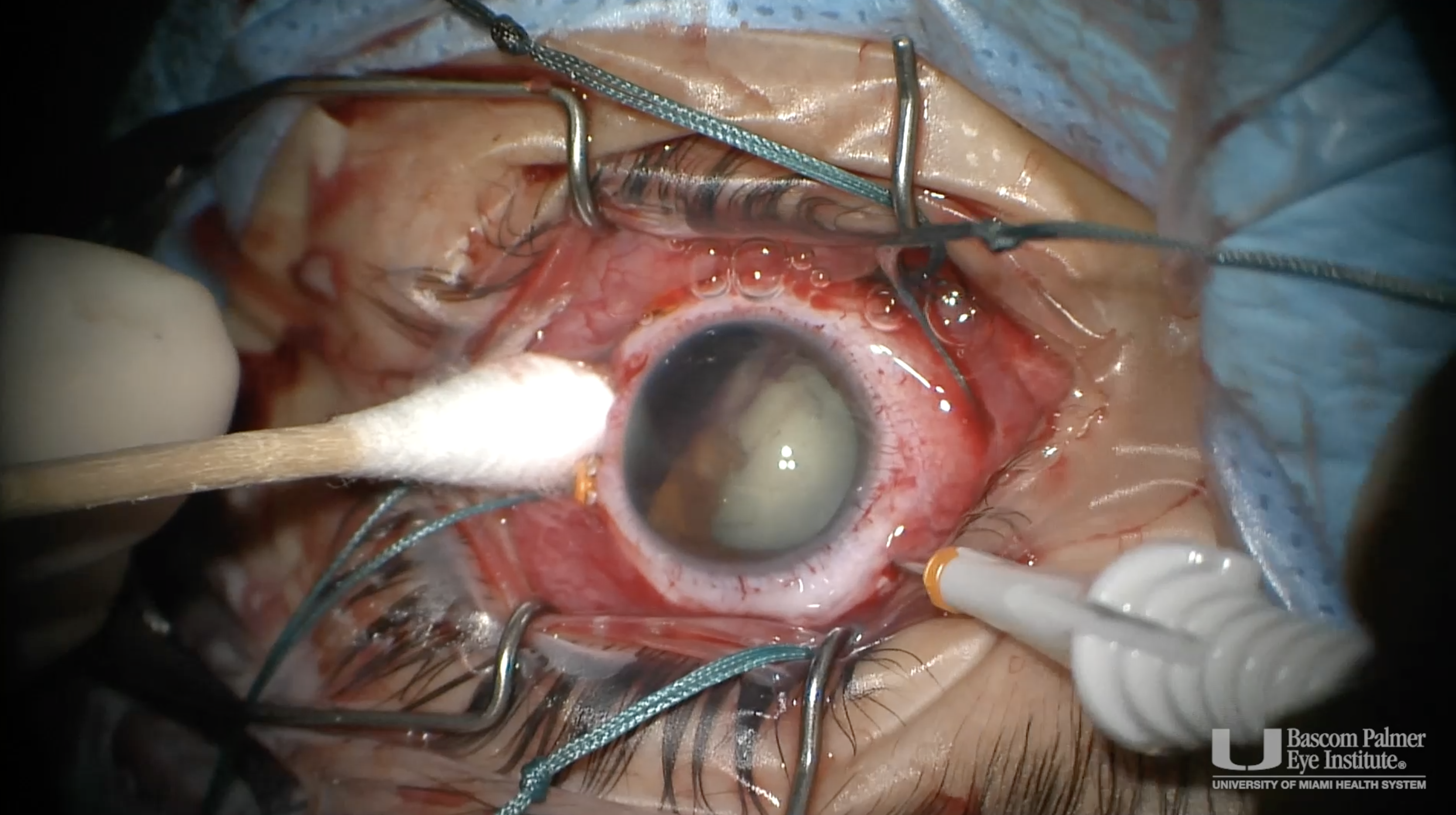
Vitrectomy, Membrane Peel and Lensectomy for Traumatic Macular Hole and Subretinal Hemorrhage due to Air Gun Injury
Specialty:
Retina and Vitreous Diseases
Type
:
Surgical Videos
Include in Catalogue?
:
Yes
Original Contributor(s)
:
Diana M. Laura, MD
Giant Retinal Tears
Specialty:
Retina and Vitreous Diseases
Type
:
Other Lectures
Include in Catalogue?
:
Yes
Presenter(s)
:
Harry W. Flynn, Jr., MD
Pneumatic Retinopexy
Specialty:
Retina and Vitreous Diseases
Type
:
Other Lectures
Include in Catalogue?
:
Yes
Presenter(s)
:
Harry W. Flynn, Jr., MD
Lamellar Keratectomy, Corneal Patch Graft, Sutured Amniotic Membrane for Neurotrophic Corneal Ulcer with Descemetocele
Specialty:
Corneal and External Diseases
Type
:
Surgical Videos
Include in Catalogue?
:
Yes
Original Contributor(s)
:
Paula W. Feng, MD; Rahul Singh Tonk, MD
Neurotrophic Corneal Ulcer
Specialty:
Corneal and External Diseases
Type
:
Ophthalmic Images
Include in Catalogue?
:
Yes
Original Contributor(s)
:
Paula W. Feng, MD; Rahul Singh Tonk, MD
Cicatricial Conjunctivitis, Steven Johnson Syndrome
Specialty:
Corneal and External Diseases
Type
:
Ophthalmic Images
Include in Catalogue?
:
Yes
Original Contributor(s)
:
Guillermo Amescua, MD; Victor H. Banda, MD
Imagining Uveitis
Specialty:
Uveitis
Type
:
Other Lectures
Include in Catalogue?
:
Yes
Presenter(s)
:
Janet L. Davis, MD
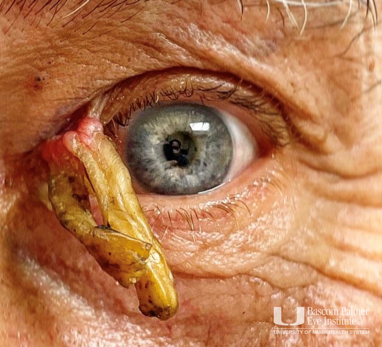
Ophthalmia Nodosa, Right Eye, Secondary to Presumed Moth Setae in the Anterior Vitreous
Specialty:
Corneal and External Diseases
Type
:
Ophthalmic Images
Include in Catalogue?
:
Yes
Original Contributor(s)
:
Noy Ashkenazy, MD, MS; Janet L. Davis, MD
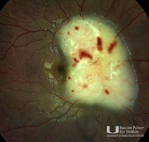
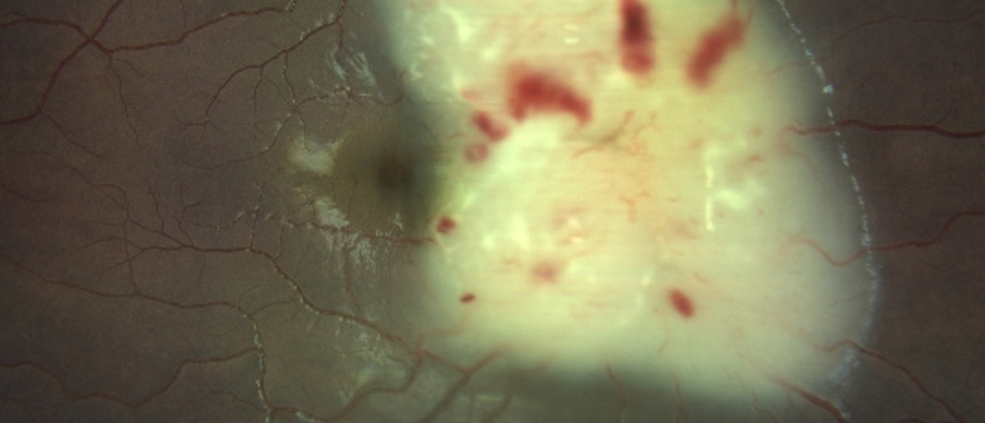
Coats' Plus Disease
Specialty:
Pediatric Ophthalmology and Strabismus
Type
:
Online Grand Rounds
Include in Catalogue?
:
Yes
Presenter(s)
:
Maria Paula Fernandez, MD
Faculty Discussant(s)
:
Audina M. Berrocal, MD
«
Previous page
1
Page 1
…
49
Page 49
50
Page 50
51
Page 51
52
Page 52
53
Page 53
54
Page 54
55
Page 55
56
Page 56
57
Page 57
58
Page 58
…
65
Page 65
»
Next page
Medical Disclaimer
|
Terms of Use
|
Privacy Statement
|
HIPAA Notice of Privacy Practices
|
About Us
|
FAQs
© 2026 University of Miami Health Systems. All Rights Reserved.