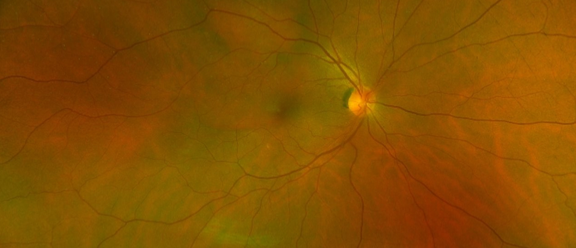
Circumscribed Choroidal Hemangioma
A patient presented with blurry vision OD. VA 20/40 OD and 20/20 OS. DFE significant for elevation along the superior arcade. Imaging workup significant for OCT showing foveal involving serous retinal detachment OD. FA and ICG showed hyperfluoroescent lesion superiorly. 12x12 SSOCTA showed elevation corresponding with areas of leakage on the FA/ICG. B-Scan showed area of thickening in posterior pole with high internal reflectivity. The patient underwent PDT and 6 weeks after treatment imaging noted resolution of the SRF, mass elevation and improvement in vision. The treatment effect was sustained at three months post procedure.
Presentation Date: 03/11/2021
Issue Date: 04/02/2021
Please log in or click on ENROLL ME to access this course.
