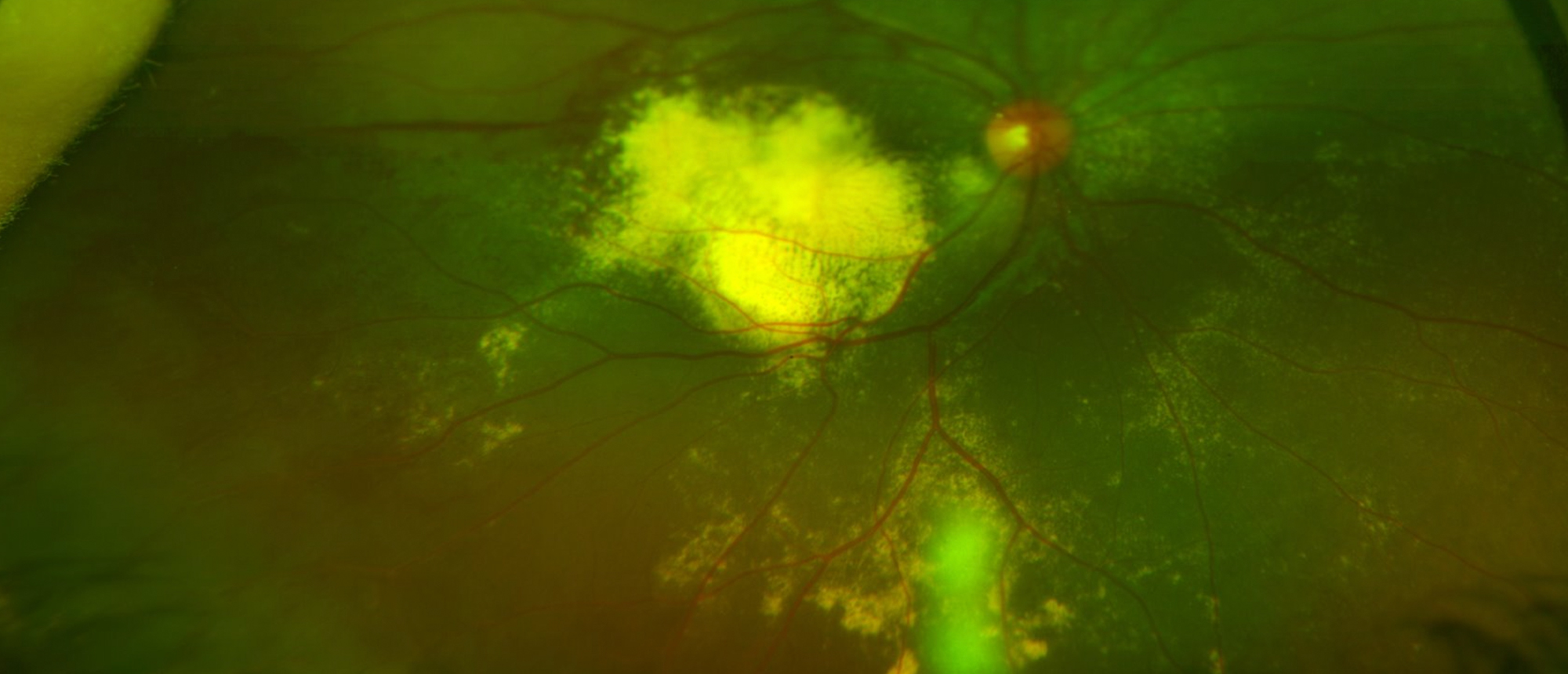
Coats' Disease with Secondary Pseudoangiomatous Acquired Retinal Gliosis
A healthy 6-year-old female was referred after she failed her school vision screening in the right eye. Exam revealed a visual acuity of count fingers (CF) at 1 foot in the right eye. Slit lamp examination was unremarkable. Fundus examination revealed peripheral telangiectasias, bulb-like aneurysms, and macular and peripheral subretinal lipid exudation, as well as a well-circumscribed peripheral orange-red elevated lesion with overlying abnormal vasculature adjacent to the ora serrata inferotemporally. She underwent exam under anesthesia in the operating room. Ocular coherence tomography revealed a large amount of macular subretinal exudation. Fluorescein angiography highlighted the abnormal vasculature seen on fundus exam and revealed lake leakage throughout the periphery. B-scan showed a homogenously hyperechoic slightly elevated retinal mass with an adjacent low-lying retinal detachment. A diagnosis of Coats’ disease with secondary pseudoangiomatous acquired retinal gliosis (PARG) was made. The patient was initially treated with intravitreal bevacizumab injection, laser, and sub-Tenon injection of triamcinolone. She had interval worsening in subretinal exudation and subsequently underwent multiple repeat treatments with improvement in exudation, resolution of the retinal detachment, and involution of the PARG lesion.
Presentation Date: 10/29/2020
Issue Date: 03/05/2021
Please log in or click on ENROLL ME to access this course.
