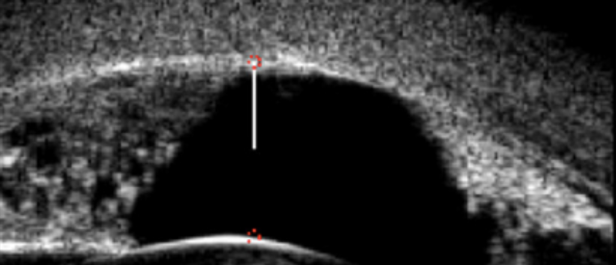
Peters Anomaly Type I
A 2-week-old girl presented with bilateral Type I Peters anomaly consisting of bilateral dense central corneal opacities and iridocorneal adhesions. Upon presentation patient had elevated IOP in both eyes (OU) (38 mmHg in the right eye (OD) and 40 mmHg in the left eye (OS)), with no other signs of glaucoma. She underwent bilateral ab-externo circumferential trabeculotomy, simultaneous lysis of iridocorneal adhesions at the time of trabecular cleavage, and optical iridectomy. This novel approach is described as the “3 in1” technique. Postoperative high-resolution ultrasound biomicroscopy (UBM) images show resolution of iridocorneal adhesions and decrease in central corneal thickness suggesting improvement in corneal edema. At the 16-month follow up visit, central corneal opacity continued to improve OU and IOP was stable OU (17 mmHg OD and 19 mmHg OS) measured by ICare rebound tonometry (ICare TAO1i, Helsinki, Finland) on no glaucoma medications.
Presentation Date: 03/12/2020
Issue Date: 08/01/2020
Please log in or click on ENROLL ME to access this course.
