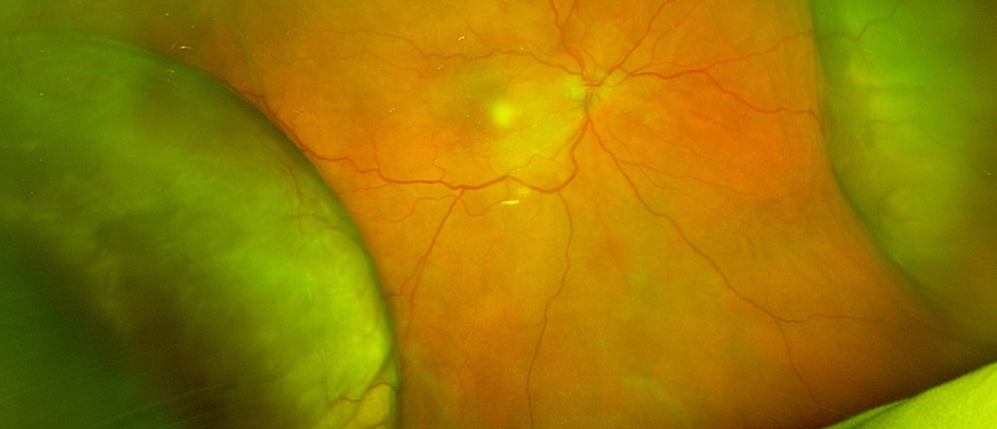
Uveal Effusion Syndrome
A patient was referred by an outside provider with a history of decreased vision in the right eye for one year. Their ocular history included glaucoma in both eyes and Fuch’s corneal dystrophy. The visual acuity was 20/200 OD and 20/30 OS. The IOP was 18 OD and 24 OS. The pupils and extraocular movements were normal. The anterior exam was notable for 1+ anterior vitreous cell in the right eye but otherwise showed findings consistent with the patient’s known prior ocular history. Dilated fundus exam revealed large choroidal effusions in all quadrants in the right eye. B-scan confirmed the choroidal detachments and showed evidence of choroidal thickening but no retinal tears, retinal detachments, masses or suspicious lesions. The left eye appeared normal. Given these findings, along with the lack of pain and inflammation, the presumed diagnosis was uveal effusion syndrome. The patient underwent scleral window surgery in the right eye. Over the next several months, the choroidal effusions resolved. By post-operative month nine, the vision was improved to 20/150. The patient has not had any recurrence of the effusion since their last follow-up.
Presentation Date: 09/07/2023
Issue Date: 09/22/2023
Please log in or click on ENROLL ME to access this course.
