Multiple Evanescent White Dot Syndrome (MEWDS)
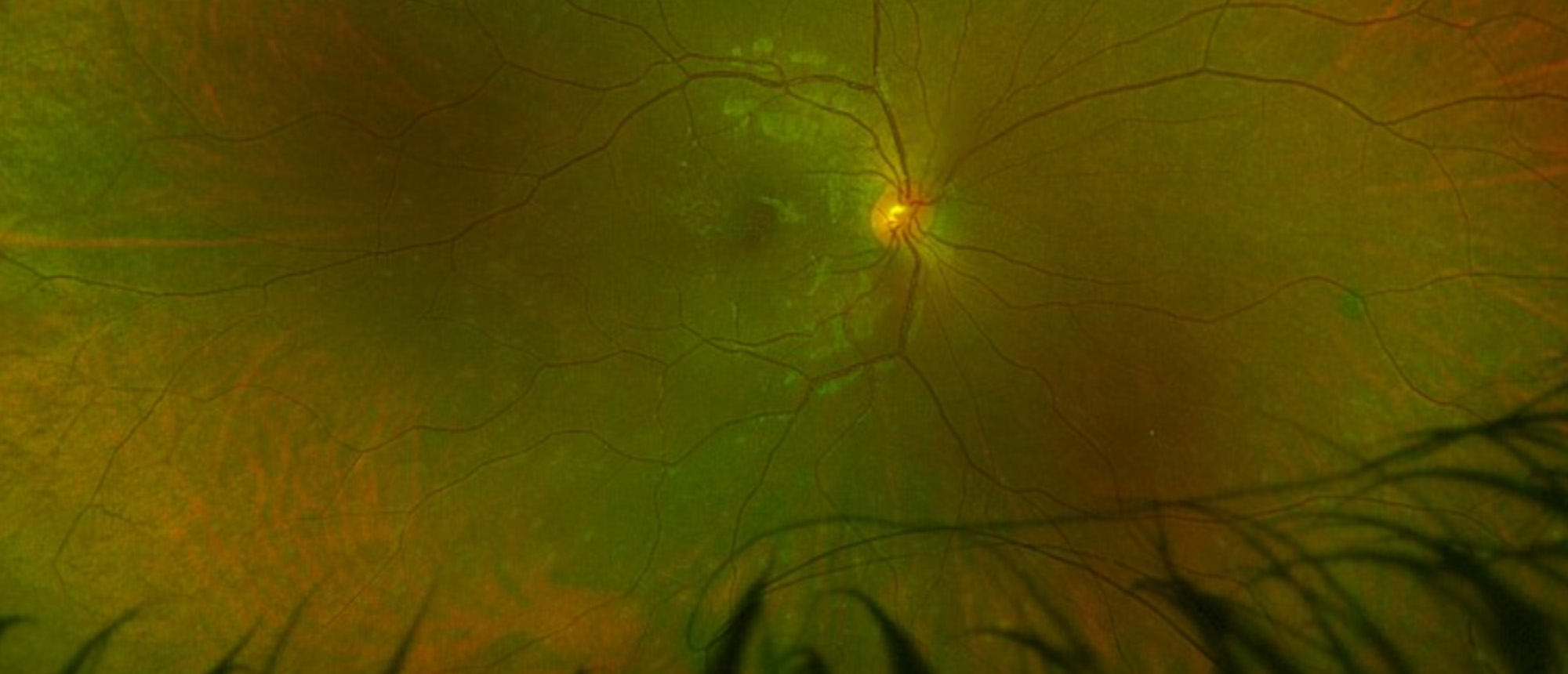
An otherwise healthy young patient without significant medical or ocular history presents with 3 days of acute-onset decreased vision in the right eye. On presentation, patient’s exam was notable for visual acuity of 20/50 in the right eye and 20/20 in the left eye. Intraocular pressure was 15 and 19 in the right and left eye, respectively. Slit lamp exam was unremarkable. Fundus exam was notable for presence of whitish-yellow spots and dots in the posterior pole and mid-periphery. Imaging was performed including fundus photos, OCT, fundus autofluorescence, fluorescein angiography and indocyanine green studies in both eyes. Imaging revealed hyperautofluorescent lesions, wreath-like hyperfluorescence around the macula, and ellipsoid zone disruption in the right eye only. Lab workup including CBC, CMP, RPR/FTA-Abs, ACE were normal. Patient improved without treatment over a course of 12 weeks, achieving baseline VA. No complications in clinical course were noted. Based on the clinical presentation, course, and imaging findings, a white dot syndrome was diagnosed.
Presentation Date: 05/18/2023
Issue Date: 06/16/2023
Please log in or click on ENROLL ME to access this course.