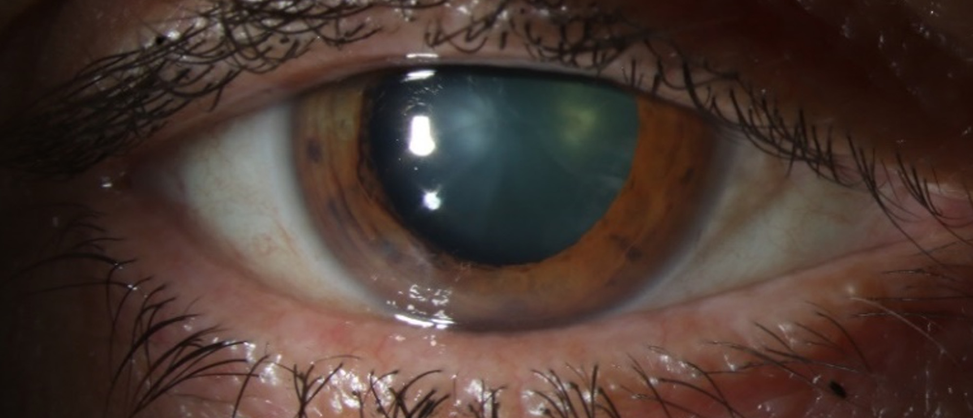
Conjunctival Lymphoma
A patient presented to the Bascom Palmer Emergency Room with a complaint of a drooping right eyelid that had been slowly progressive over 9 months. Her vision was 20/25 OD and 20/20 OS. Intraocular pressures were 17 OD and 15 OS. External exam was notable for mild right eye ptosis, proptosis, and hypoglobus. Slit lamp exam was unremarkable apart from bilateral salmon-pink colored lesions in the superior fornices, right more prominent than the left. High-resolution anterior segment OCT showed a hyporeflective, subepithelial mass in both eyes, and subsequent biopsy and pathology confirmed low-grade B-cell lymphoma. Over the next couple weeks, the patient underwent additional systemic workup. CT chest/abdomen/pelvis demonstrated enlarged lymph nodes in the bilateral axillary region and an enlarged conglomerate of lymph nodes in the pelvis, as well as nodular densities in both lungs suspicious for lymphomatous deposits. Subsequent FDG PET/CT demonstrated extensive soft-tissue density lesions above and below the diaphragm, with the largest lesion in the pelvis measuring up to 14.6 cm x 15.1 cm, and multiple soft tissue nodular lesions in the lungs. Bone marrow biopsy showed a cellular marrow with maturing trilineage hematopoiesis and a focally B-cell predominant lymphoid infiltrate that may represent involvement by B-cell lymphoma (PCR studies pending). The patient underwent IR guided biopsy of the large pelvis mass, and results of this were pending at the time of this presentation.
Presentation Date: 01/19/2023
Issue Date: 01/27/2023
Please log in or click on ENROLL ME to access this course.
