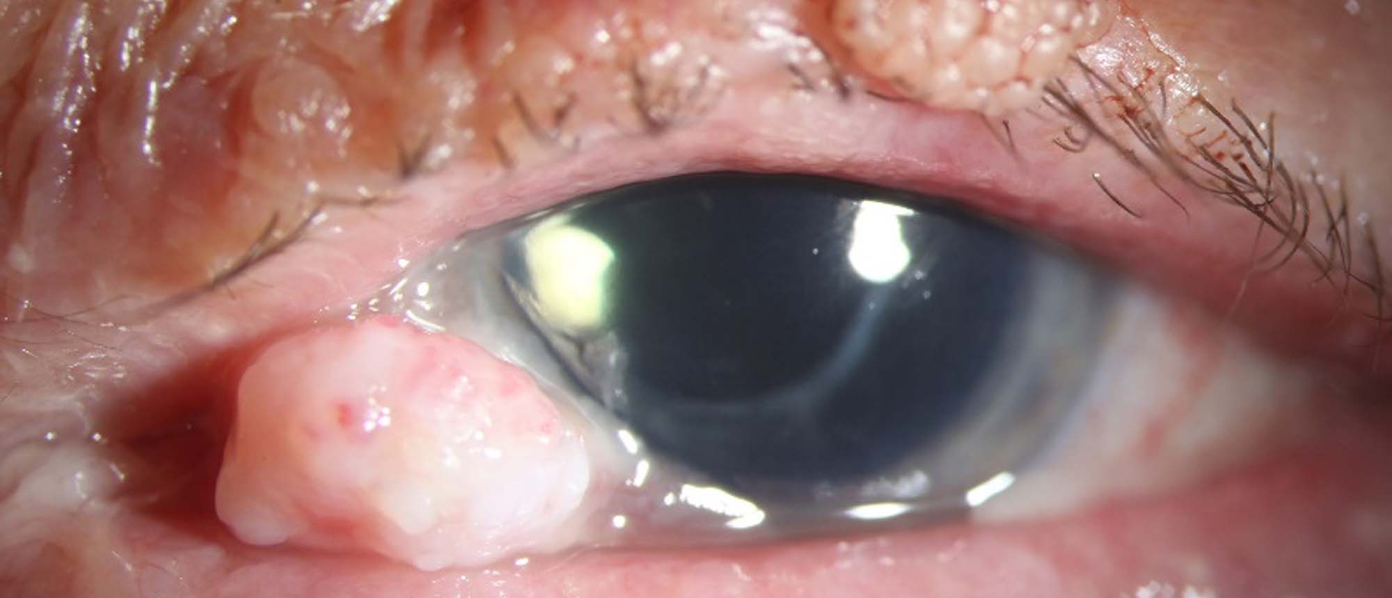
Conjunctival Squamous Cell Carcinoma, Keratoacanthoma Type
A patient presented with a rapidly enlarging lesion of the nasal conjunctiva of the left eye. The past ocular history was significant only for bilateral surgical removal of cataracts with intraocular lens placement. The patient had a past medical history notable for renal cell carcinoma (RCC). Upon presentation, slit lamp examination disclosed a white, dome-shaped, pedunculated, nodular mass with an irregular surface and minimal vasculature attached to the perilimbal area of inferonasal conjunctiva of the left eye. Exam also demonstrated an unremarkable anterior segment with posterior chamber intraocular lenses in both eyes. Anterior segment optical coherence tomography (OCT) of the left eye demonstrated a thickened, hyperreflective epithelium with an abrupt transition from normal to abnormal epithelium, as well as shadowing over a disorganized subepithelial lesion. High Resolution Ultrasound B-scan did not demonstrate scleral extension of the lesion. A date was set for tissue biopsy, however the patient’s lesion underwent spontaneous involution prior to biopsy completion. The patient underwent surgical excision of remaining lesion with a no touch technique and wide 4mm surgical margins. Cryotherapy was applied to the conjunctival margins as well as the scleral bed. Amniotic membrane transplantation (AMT) was placed with fibrin glue. Surgical histopathology was significant for mucosal epithelium with faulty epithelial maturational sequencing extending full thickness. The tissue was acanthotic with marked parakeratosis presenting in a “cup-like” or “dome like” configuration. These findings were consistent with squamous cell carcinoma, keratoacanthoma subtype. Post-operative anterior segment OCT showed intact epithelium, with no mass lesion. The patient completed two cycles of treatment with interferon α-2B and management of post-surgical dellen with complete resolution. 3 month follow up slit lamp examination and anterior segment OCT displayed complete resolution of the lesion without evidence of recurrence, and a normalized epithelium with some stromal thinning. There was no recurrence of the lesion at 2.5 year follow up.
Presentation Date: 09/23/2021
Issue Date: 02/25/2022
Please log in or click on ENROLL ME to access this course.

