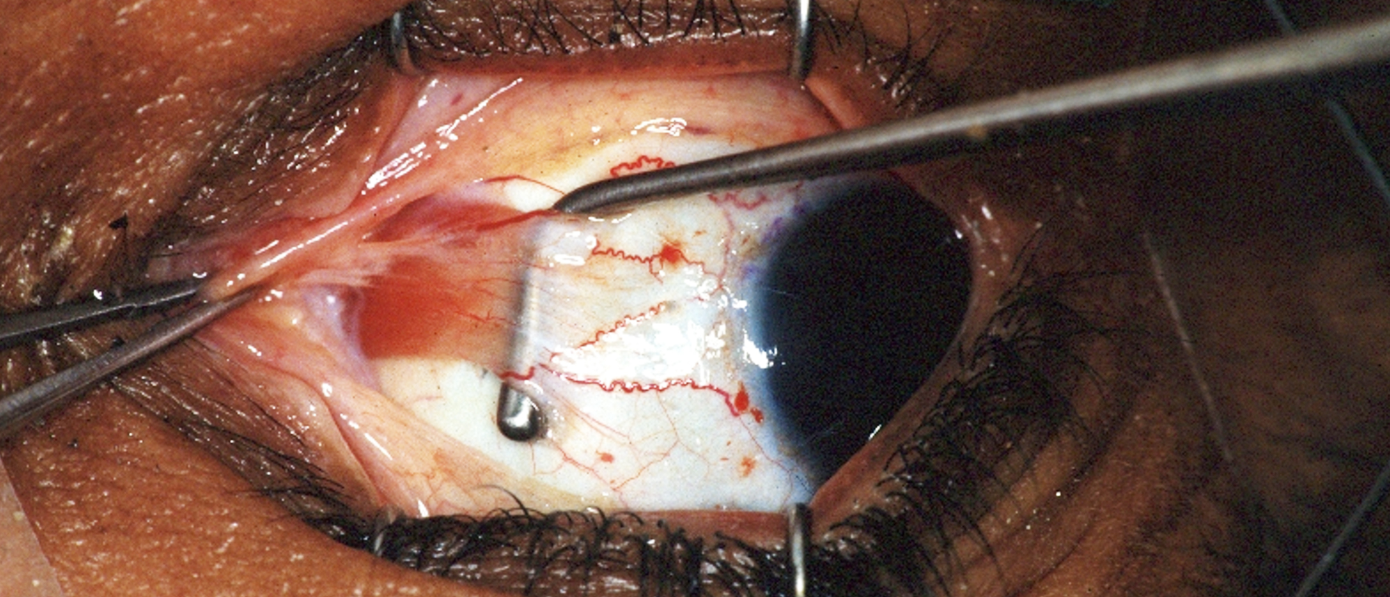
Idiopathic Intracranial Hypertension
A 39-year-old male presented to the BPEI ER with blurry vision in his right eye for 2 weeks. Examination revealed CF OD, 20/60 OS vision, right APD, and bilateral disc edema. OCT showed diffusely elevated RNFL thickness and HVF 30-2 was not able to be performed on the right eye but demonstrated an inferior arcuate defect in the left eye. CT scan revealed a partially empty sella. The patient was presumed to have idiopathic intracranial hypertension and a right optic nerve sheath fenestration was performed to prevent progression of vision loss.
Presentation Date: 10/15/2020
Issue Date: 03/05/2021
Please log in or click on ENROLL ME to access this course.
Include in Catalogue?: No
Vetted By: David T. Tse, MD
Vetted Date: May 28, 2025
Presenter(s): Michelle M. Maeng, MD
Faculty Discussant(s): David T. Tse, MD

