Cranial Nerve 6 Palsy from Metastatic Cholangiocarcinoma
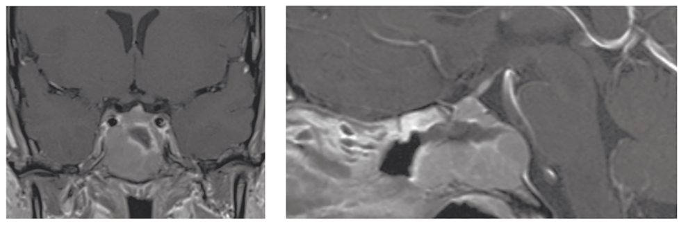
A 34-year-old woman presented with 2 weeks of diplopia. The patient had binocular horizontal diplopia with a right cranial nerve 6 palsy and moderate-to-severe headache of 1-week duration. Magnetic resonance imaging (MRI) of the brain and orbits revealed
a 2.2 x 3.0 x 2.5 cm sellar/suprasellar mass with internal cystic necrotic component and abutment of the cavernous sinuses. Pathology of the clival lesion revealed a moderately differentiated adenocarcinoma consistent with cholangiocarcinoma. Positron
emission tomography/computed tomography (PET/CT) scan demonstrated a large hypermetabolic partially necrotic mass in the right hepatic lobe extending to the caudate lobe. Next generation RNA whole transcriptome sequencing of the metastatic tissue
revealed an FGFR2-BICC1 gene fusion. Gross total resection was achieved via an endoscopic transnasal transsphenoidal approach with resolution of diplopia. The mass recurred 3 weeks later, with new onset diplopia. The patient received stereotactic
radiotherapy applied to the brain lesion and began treatment with gemcitabine and cisplatin and was alive at 7 months after initial presentation.
Presentation Date: 07/23/2020
Issue Date: 01/22/2021
Please log in or click on ENROLL ME to access this course.

