
Atypical Peripheral Exudative Hemorrhagic Chorioretinopathy (PEHCR)
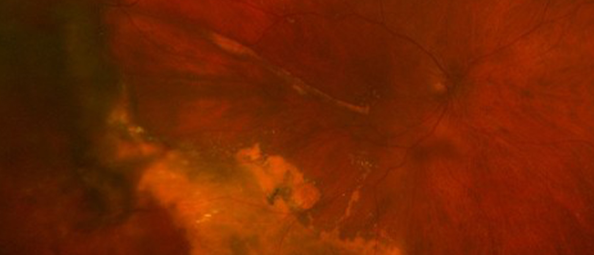
Peripheral exudative hemorrhagic chorioretinopathy (PEHCR) is an uncommon degenerative chorioretinal disorder that can present as an elevated peripheral mass, mimicking choroidal melanoma. We present a patient referred for decreased vision and retinal hemorrhage following laser peripheral iridotomy (LPI). Widefield imaging revealed a large elevated inferotemporal lesion with subretinal fibrosis, lipid exudation, and hemorrhage extending toward the macula. OCT demonstrated subretinal fluid extending to the fovea, while B-scan showed a plateau-shaped lesion with heterogeneous reflectivity and a possible clot retraction cleft. Fluorescein angiogram (FA) and Indocyanine Green angiography (ICG) revealed no evidence of neo-vascularization. Multimodal imaging supported a diagnosis of atypical PEHCR, with polypoidal choroidal vasculopathy and choroidal melanoma among the differential diagnoses. Intravitreal bevacizumab was administered for macular involvement with resolution of sub-retinal fluid and improvement in BCVA. Although observation is appropriate for many PEHCR lesions, anti-VEGF therapy may be considered when fluid or hemorrhage threatens the macula. Posterior segment complications following LPI have been reported; however, an association with PEHCR has not been established. This case highlights the importance of multimodal imaging in distinguishing PEHCR from other etiologies and raises the question of a potential temporal association between LPI and the development or recognition of peripheral choroidal pathology.
Presentation Date: 11/12/2026
Issue Date: 11/13/2026
Please log in or click on ENROLL ME to access this course.
