
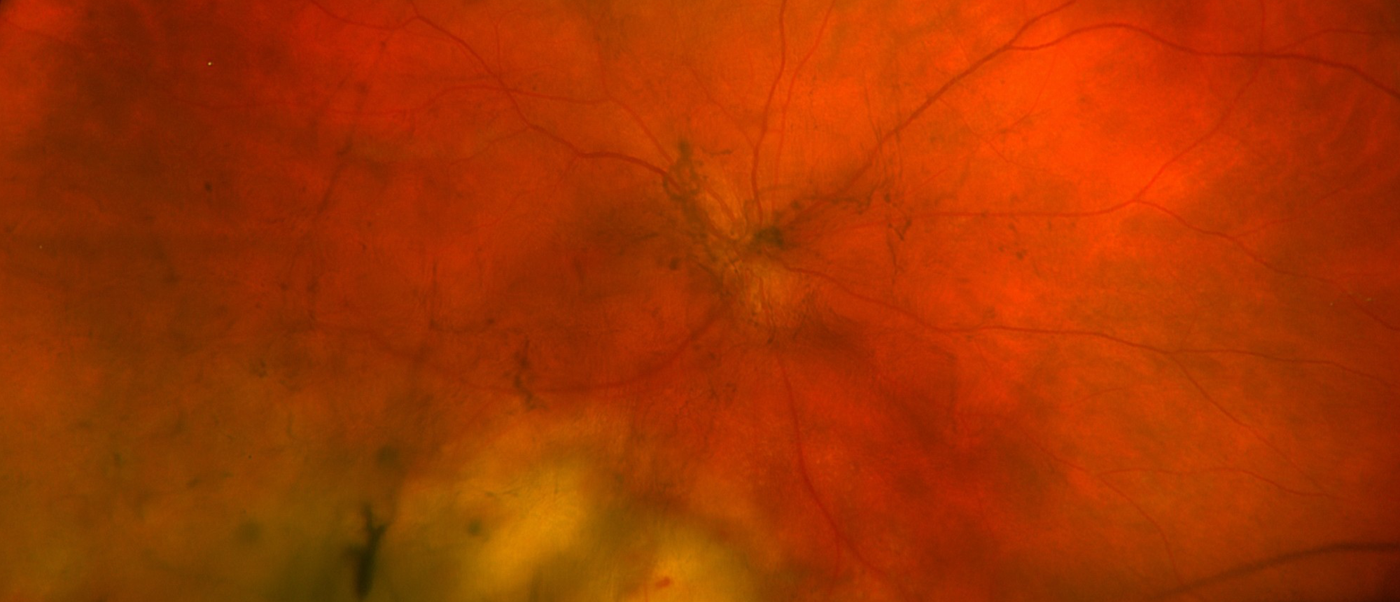
Endogenous Endophthalmitis with Subretinal Abscess Likely Secondary to Dental Infection
A patient with coronary artery disease status post multiple coronary interventions, ischemic cardiomyopathy, prior stroke, and newly identified diabetes presented with four days of right eye pain, blurred vision, and photopsias beginning approximately three weeks after dental work. After two weeks of topical corticosteroids at an outside clinic, his optometrist noted optic nerve edema and referred him to a community emergency department to “rule out giant cell arteritis and stroke.” Inflammatory markers, NIH stroke scale, CT angiography, and MRI of the brain and orbits were all normal; because ophthalmology was unavailable, emergent transfer was recommended, but the patient declined and left against medical advice. Four days later, dilated examination at the Bascom Palmer Eye Institute revealed anterior chamber cell, vitritis, and an inferotemporal milky-white subretinal lesion with overlying hemorrhages consistent with endogenous endophthalmitis with subretinal abscess. He underwent vitreous tap with intravitreal vancomycin, ceftazidime, and voriconazole, repeated one week later, and was admitted for systemic antibiotics. An exhaustive source evaluation — blood and vitreous cultures, HIV, syphilis and tuberculosis testing, toxoplasma serologies and PCR, computed tomography of the chest, abdomen, and pelvis, and echocardiography — was entirely negative, leaving recent dental bacteremia as the presumed source. On prolonged systemic therapy the abscess progressively consolidated and vision recovered from 20/100 to 20/50 without vitrectomy. This case highlights endogenous endophthalmitis masquerading as neuro-ophthalmic disease, the association between dental procedures and hematogenous ocular infection, the management of culture-negative disease, and transfer obligations when subspecialty care is unavailable.
Presentation Date: 08/27/2026
Issue Date: 08/28/2026
Please log in or click on ENROLL ME to access this course.
