
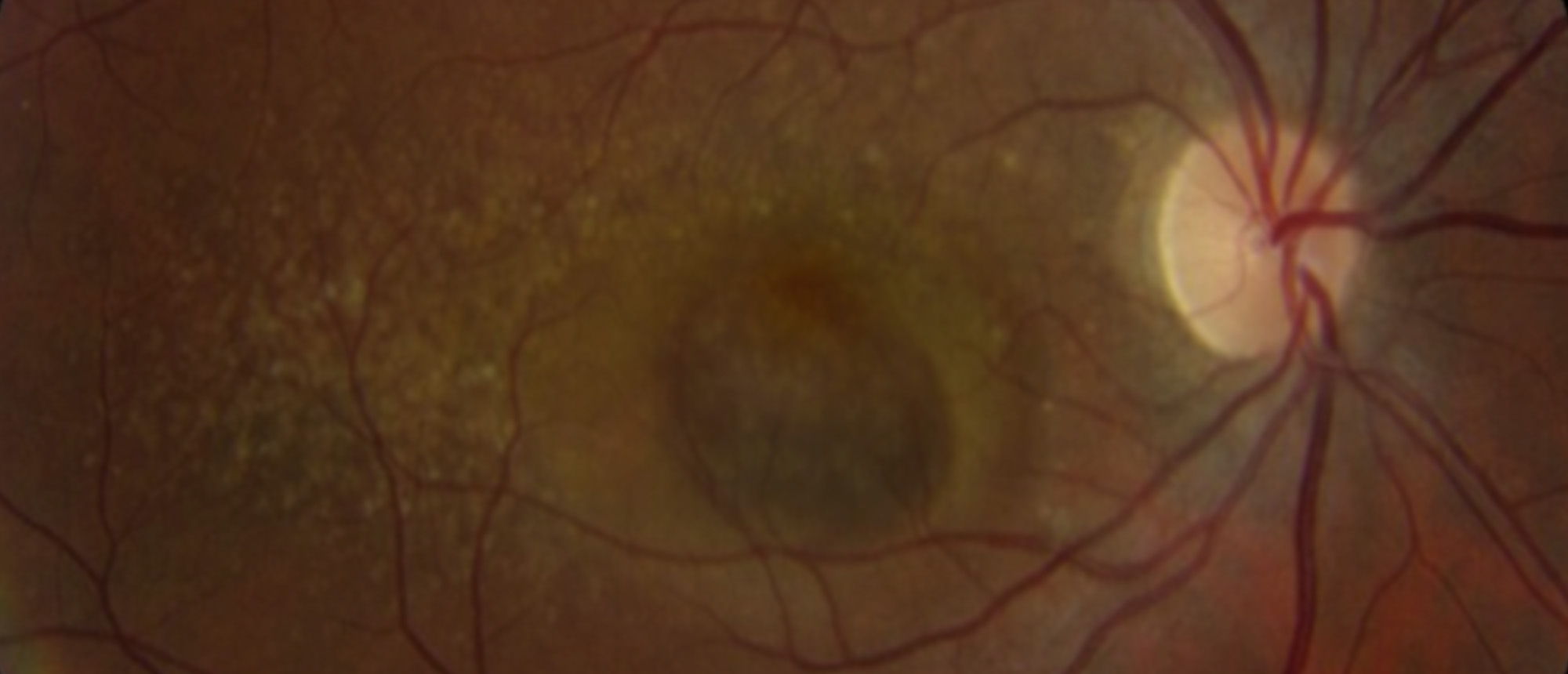
Polypoidal Choroidal Vasculopathy
A patient was referred by an outside provider for decreased vision in the right eye. The exam was notable for large hemorrhagic PED in the right eye and numerous drusen in both eyes. OCT showed large PED with SRF in the right eye. ICGA demonstrated polyps in the right eye. The patient was subsequently diagnosed with PCV and treated with intravitreal anti-VGEF. SSOCTA imaging showed the polyps in this patient and other PCV cases were indeed a variant of type 1 MNV and can evolve into classic type 1 MNV after anti-VEGF treatment.
Presentation Date: 04/16/2026
Issue Date: 04/24/2026
Please log in or click on ENROLL ME to access this course.
Include in Catalogue?: No
Presenter(s): Da Meng, MD, PhD
Faculty Discussant(s): Philip J. Rosenfeld, MD, PhD
