
Abstract
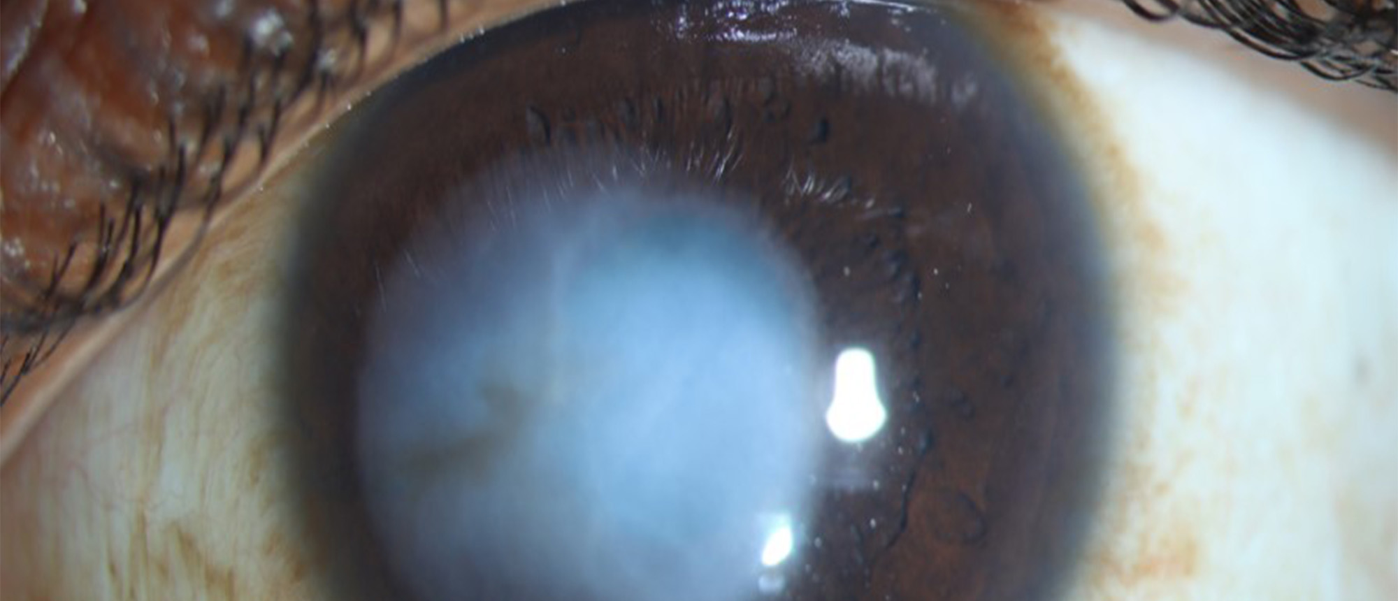
A patient with a past ocular history of infectious keratitis in the left eye presents to the clinic for vision rehabilitation. He reported a decreased visual acuity in the left eye that never recovered after the infection. Her exam was notable for visual acuity of 20/20 in the right eye and 20/70 best corrected in the left eye. Intraocular pressure was 14 and 17. Slit lamp exam was most notable for a central dense corneal scar measuring 6x6 mm involving the visual axis. The anterior segment-OCT confirms the involvement of the anterior 350 microns of the cornea, with a relatively preserved posterior 1/3 of the stroma. Due to severe intolerance, the patient was not using Scleral contact lenses and wanted to avoid long-term immunosuppression. The scarred tissue was extending too deeply in the corneal stroma to try the phototherapeutic keratectomy option without the risk of corneal ectasia. The patient then underwent a femtosecond laser-assisted Anterior Lamellar Keratoplasty in the left eye. The two-step procedure involved the creation of a donor graft from a whole globe mounted on a custom-made globe holder under a femtosecond laser device. Subsequently, the same laser was used to remove the recipient tissue, the donor graft was positioned on the recipient area and secured in place by drying the flap edges and by placing a bandage contact lens. After the first week postop, the contact lens was removed, and the graft was fully epithelialized. Best corrected visual acuity on the left eye was significantly improved to 20/40.
Presentation Date: 05/15/2025
Issue Date: 04/17/2026
Include in Catalogue?: No
Presenter(s): Francesco Pozzo Giuffrida, MD, PhD
Faculty Discussant(s): Florence A. Cabot, MD
