
Congenital Corneal Keloid
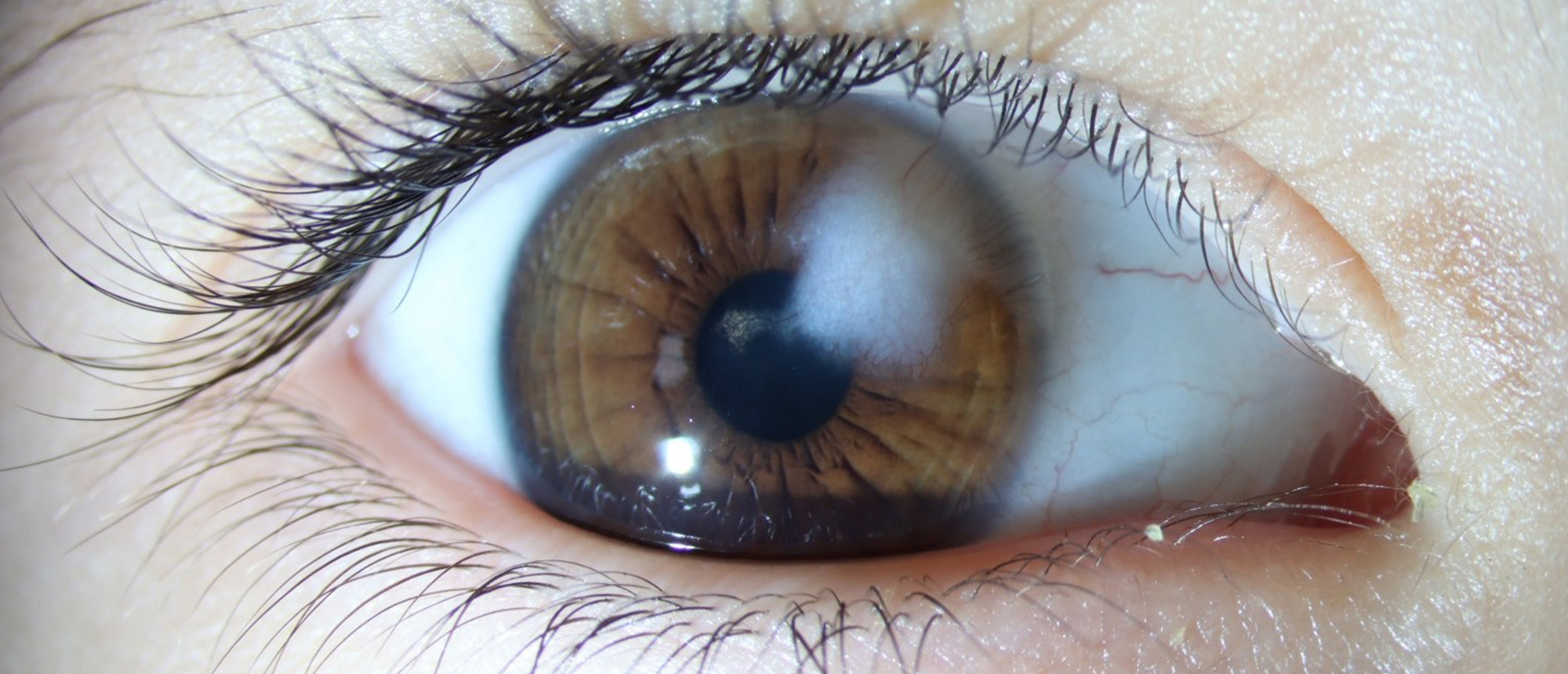
A patient was brought to clinic by his mother for a white spot on the right eye. The patient was born full-term with no complications. They had a history of delayed speech but was otherwise meeting all developmental milestones. The family history was unremarkable. The vision was 20/100 for the right eye and 20/20 for the left eye. The intraocular pressures were 14 OD and 13 OS. The patient had limited stereoscopy. Slit lamp exam demonstrated a paracentral 5x5mm white, slightly raised, subepithelial corneal lesion. The remainder of the anterior and posterior exam was normal in the right eye, and the left eye had no abnormal findings. Anterior segment optical coherence tomography demonstrated a hyper-reflective subepithelial lesion with approximately 20% stromal depth. On further history, the patient had developed significant anisometropic myopic astigmatism at around age four and had started treatment for amblyopia with patching and full-time glasses. Given the exam and history, the decision was made to remove the lesion. The patient was taken to the OR for superficial keratectomy with AMT and mitomycin-C. By post-operative month 1, the vision improved to 20/60 and the cornea remained clear. The patient is using topical losartan drops and continues to do well.
Presentation Date: 05/08/2025
Issue Date: 04/17/2026
