
Adult Onset Foveomacular Vitelliform Dystrophy
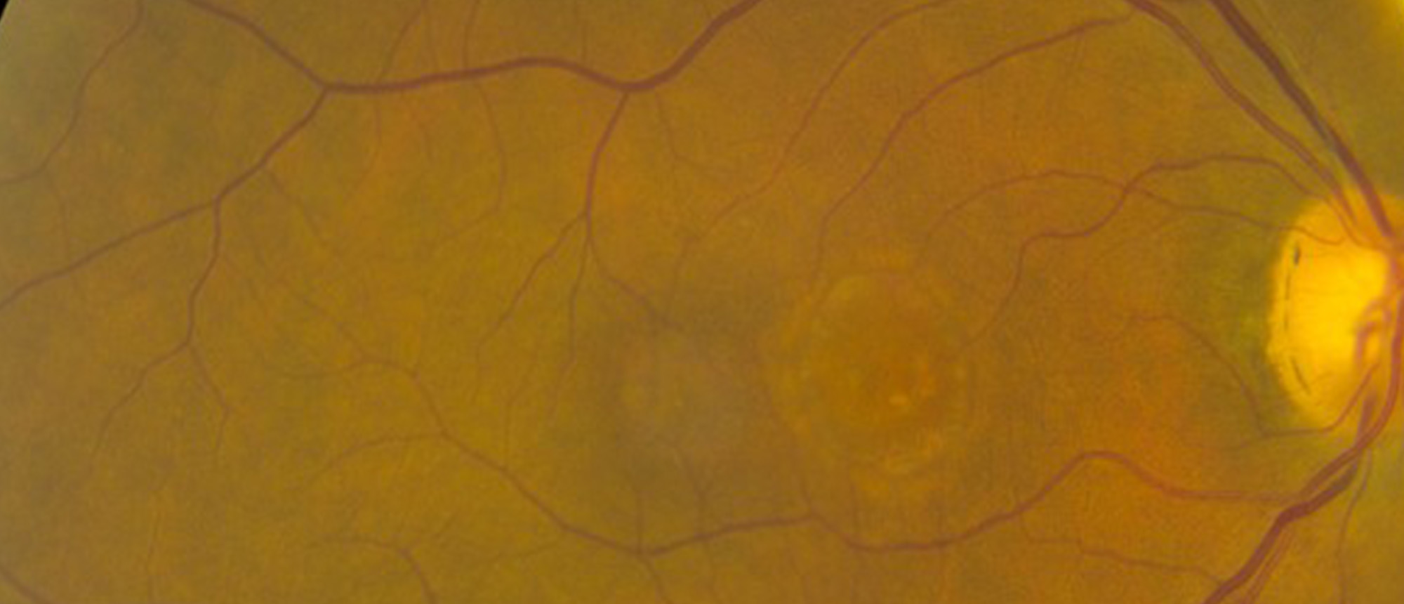
A patient presented with complaints of painless, progressive decrease in vision in both eyes. On examination, her BCVA was 20/60 in right eye and 20/100 in left eye. Fundus examination showed RPE mottling in both eyes, Optical Coherence Tomography showed Macular atrophy in the right eye and intra and subretinal fluid in left eye. OCTA showed Choroidal neovascular membrane in the left eye. Going back in history and review of her records showed bilateral yellow lesions in both eyes and OCT showed subretinal hyperreflective material in both eyes.
Conclusion: Diagnosis of AOFVD can be challenging due to overlapping symptoms and features with Best Dystrophy and Age-related macular degeneration. Multimodal imaging is crucial for monitoring the disease and early detection of complications.
Presentation Date: 05/01/2025
Issue Date: 04/17/2026
Please log in or click on ENROLL ME to access this course.
