
Extraocular Silicone Oil Deposition
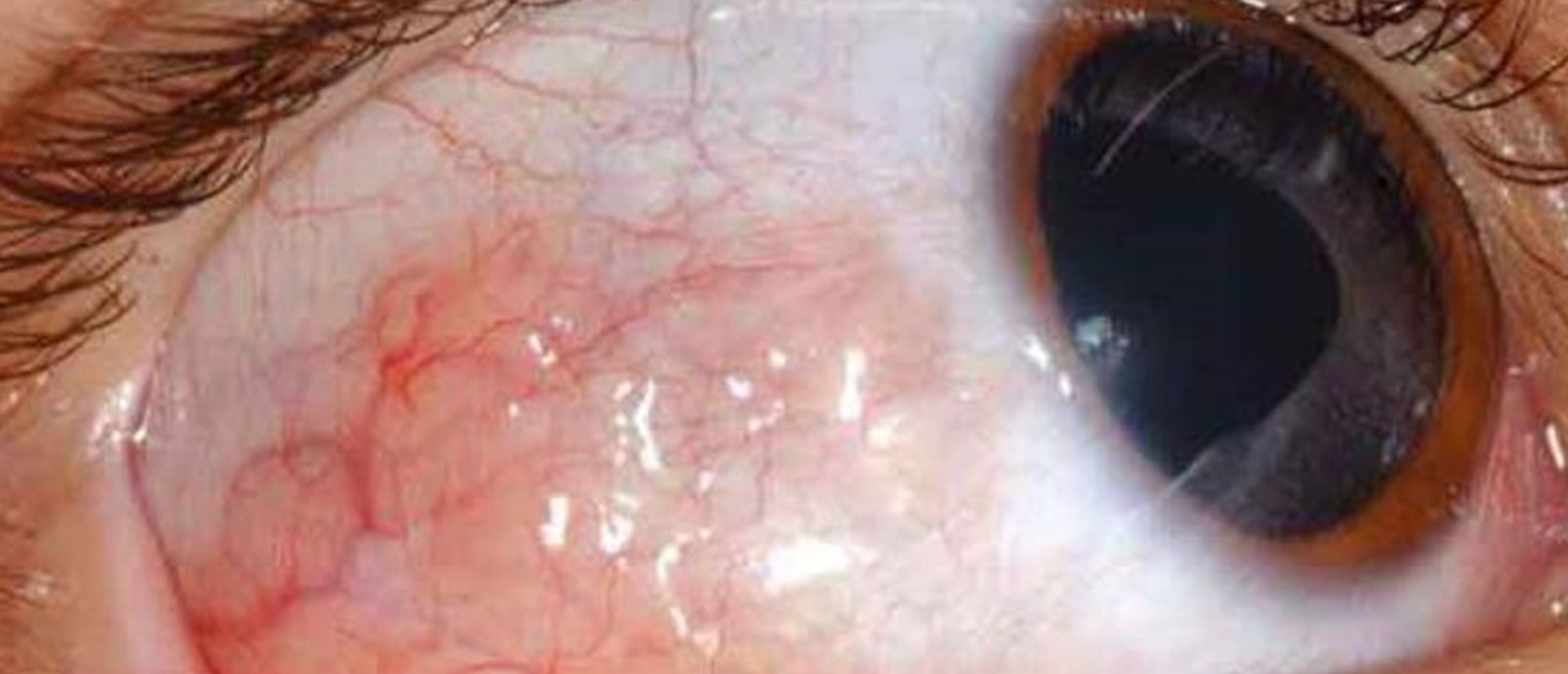
A patient presented with a painless, gradually enlarging subconjunctival cystic lesion of the right eye. Anterior segment examination revealed a translucent, cystic-appearing nasal bulbar conjunctival mass. Anterior segment optical coherence tomography (AS-OCT) demonstrated hyporeflective cystic spaces within the substantia propria, prompting excisional biopsy for definitive diagnosis. The patient’s ocular history was significant for a giant retinal tear with macula-off rhegmatogenous retinal detachment in the right eye, treated seven months prior with scleral buckle, pars plana vitrectomy (PPV), 1000-centistoke silicone oil tamponade, and subsequent silicone oil removal with fluid–air exchange and endolaser. Histopathologic evaluation of the conjunctival lesion revealed dropout spaces consistent with silicone oil vacuoles surrounded by CD68- and CD163-positive histiocytes, confirming extraocular silicone oil migration with associated chronic granulomatous inflammation and foreign body reaction. The lesion was successfully managed with surgical excision and amniotic membrane transplantation, with no recurrence. This case highlights extraocular migration of intraocular silicone oil as an uncommon but important late complication of vitreoretinal surgery. Silicone oil may migrate through sclerotomy sites or areas of scleral thinning and present months to years after surgery as conjunctival, eyelid, or orbital lesions, mimicking benign or malignant processes. Multimodal imaging, particularly AS-OCT and orbital CT, can aid in localization and surgical planning. Recognition of this entity is critical for both ophthalmologists and pathologists to avoid misdiagnosis and to guide appropriate management, as most cases are effectively treated with complete surgical excision.
Presentation Date: 02/19/2026
Issue Date: 04/03/2026
Please log in or click on ENROLL ME to access this course.
