
Bullous Central Serous Retinopathy with Choroidal Effusions
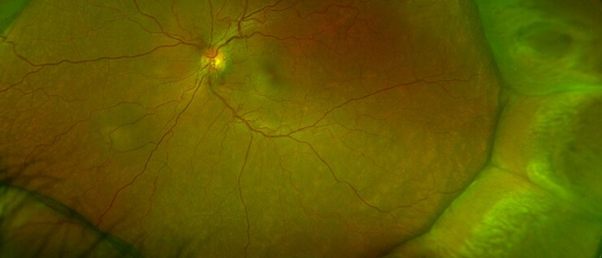
A patient with no past ocular history presents with a 1-day history of decreased vision in his left eye. Examination revealed large choroidal effusions. Further history revealed past systemic steroid and testosterone use. On multimodal imaging he had a large dome shaped serous retinal detachment with retinal pigment epithelial changes. Ocular ultrasonography showed choroidal thickening of both eyes, with remarkable effusions of the left eye. He had multiple areas of leakage on fluorescein angiography, and choroidal congestion and hyperpermeability on indocyanine green. His clinical examination, history, and findings on multimodal imaging lead to the diagnosis with central serous chorioretinopathy which presented in an atypical fashion, with choroidal effusions.
Presentation Date: 11/13/2025
Issue Date: 01/09/2026
Please log in or click on ENROLL ME to access this course.
