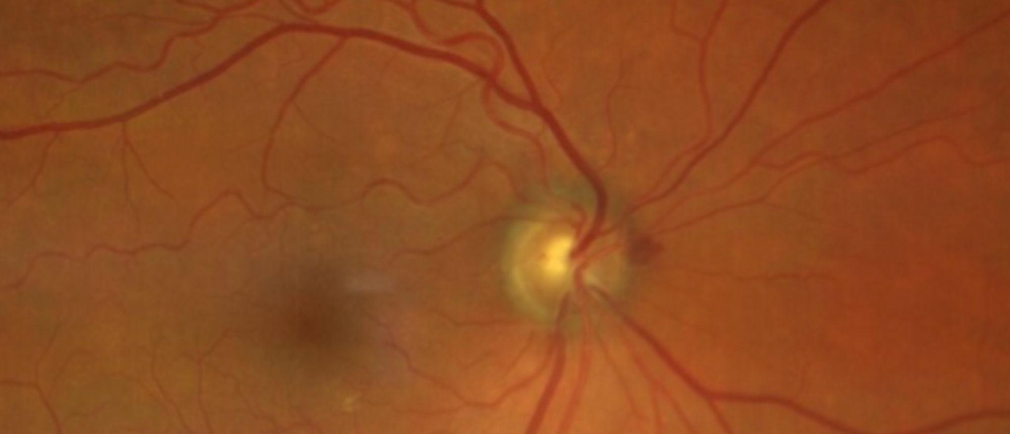
Infiltrative Optic Neuropathy Secondary to Pleomorphic Mantle Cell Lymphoma
A patient presented to the ophthalmology emergency room with a 2-day history of decreased vision of his left eye. Examination revealed vision of 20/25 OD and 20/50 OS, with a relative afferent pupillary defect in the left eye. Neuro-ophthalmologic examination revealed a constricted visual field on the left, decreased color plates to 3 out of 11 on the left, 20% red desaturation and 18% relative brightness both on the left. Dilated fundus exam showed optic nerve edema on the left. Humphry visual field showed nasal field loss greater inferiorly on the left, with the right being within normal limits. RNFL confirmed the nerve edema on the left, with an average thickness of 223 um. MRI of the brain and orbits showed mild atrophy of the right optic nerve and abnormal signal I the left optic nerve. A complete blood count showed the white blood cells to be severely elevated to 56.0. The patient was transferred to the hospital for a complete systemic workup. Bone marrow biopsy with flow cytology was consistent with Mantle cell lymphoma pleomorphic variant. A PET scan was also performed to complete staging. A lumbar puncture with flow cytometry analysis confirmed confirmed leptomeningeal involvement of the lymphoma. The patient was diagnosed with Mantle cell lymphoma with infiltrative optic neuropathy. The patient was started on both systemic and intrathecal chemotherapy.
Presentation Date: 11/07/2024
Issue Date: 11/13/2024
Please log in or click on ENROLL ME to access this course.

