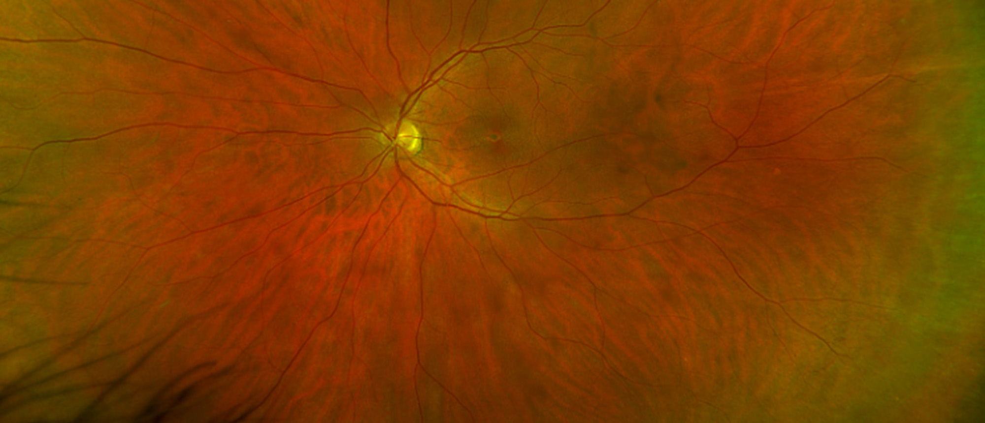
PRPH2-Associated Pattern Dystrophy
A middle-aged patient presented to the Bascom Palmer Eye Institute with central blurred vision in the left eye for 2 weeks. Past medical and ocular history were unremarkable. Past family history was notable for a mother who was diagnosed with age-related macular degeneration in her 40s and grandparents with age-related macular generation. Visual acuity in the right eye was 20/20 and in the left eye was 20/20 eccentrically. Intraocular pressure was normal in both eyes. Anterior segment exam was unremarkable in both eyes. Dilated fundus exam in both eyes were notable for bilateral foveal retinal pigment epithelium hyperplasia. Optical coherence tomography revealed small foveal RPE hyperplasia, parafoveal drusenoid PED, and foci of hyperreflectivity at the outer plexiform layer in the right eye and foveal RPE hyperplasia in the left eye. Fluorescein angiography revealed peripheral nonperfusion, mild supernumerary vessels, and mild peripheral small vessel leakage with circumferential vessels in both eyes. Fundus autofluorescence revealed parafoveal hyperautofluorescnece in both eyes with mild blocking secondary to pigmentary changes. Genetic testing revealed a heterozygous mutation in peripherin-2 (PRPH2) suggestive of a diagnosis of PRPH-2 associated pattern dystrophy. Multifocal and full-field electroretinograms were normal.
Presentation Date: 07/25/2024
Issue Date: 08/09/2024
Please log in or click on ENROLL ME to access this course.

