
Sympathetic Ophthalmitis
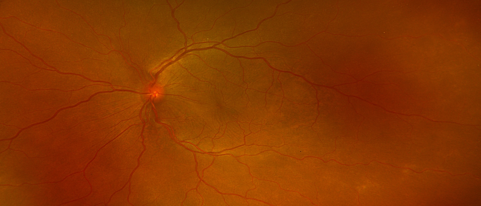
A patient presented with complaints of decreased vision in left eye of two days duration. He had a past ocular history of repair of scleral tear in the right eye 3 weeks back. On examination, his BCVA was HM in right eye and 20/300 in left eye. Anterior segment examination showed sutured scleral tear in the right eye with blood in anterior chamber. Examination of left eye showed 3+ Ac cells and 2+flare with 360-degree posterior synechiae. Fundus examination of right had poor view and left eye showed 1+ vitreous cells, yellow white chorioretinal lesion in the periphery and serous retinal detachments inferiorly. Optical Coherence Tomography showed multiple pockets of subretinal fluid, bacillary detachment and thickened choroid in left eye. Bscan showed diffuse thickening of choroid in the left eye. Diagnosis of sympathetic ophthalmia can be challenging due to overlapping symptoms and features with other noninfectious or infectious panuveitis. Meticulous history and imaging is crucial for early diagnosis and prompt treatment to restore vision.
Presentation Date: 11/13/2025
Issue Date: 07/03/2026
Please log in or click on ENROLL ME to access this course.
