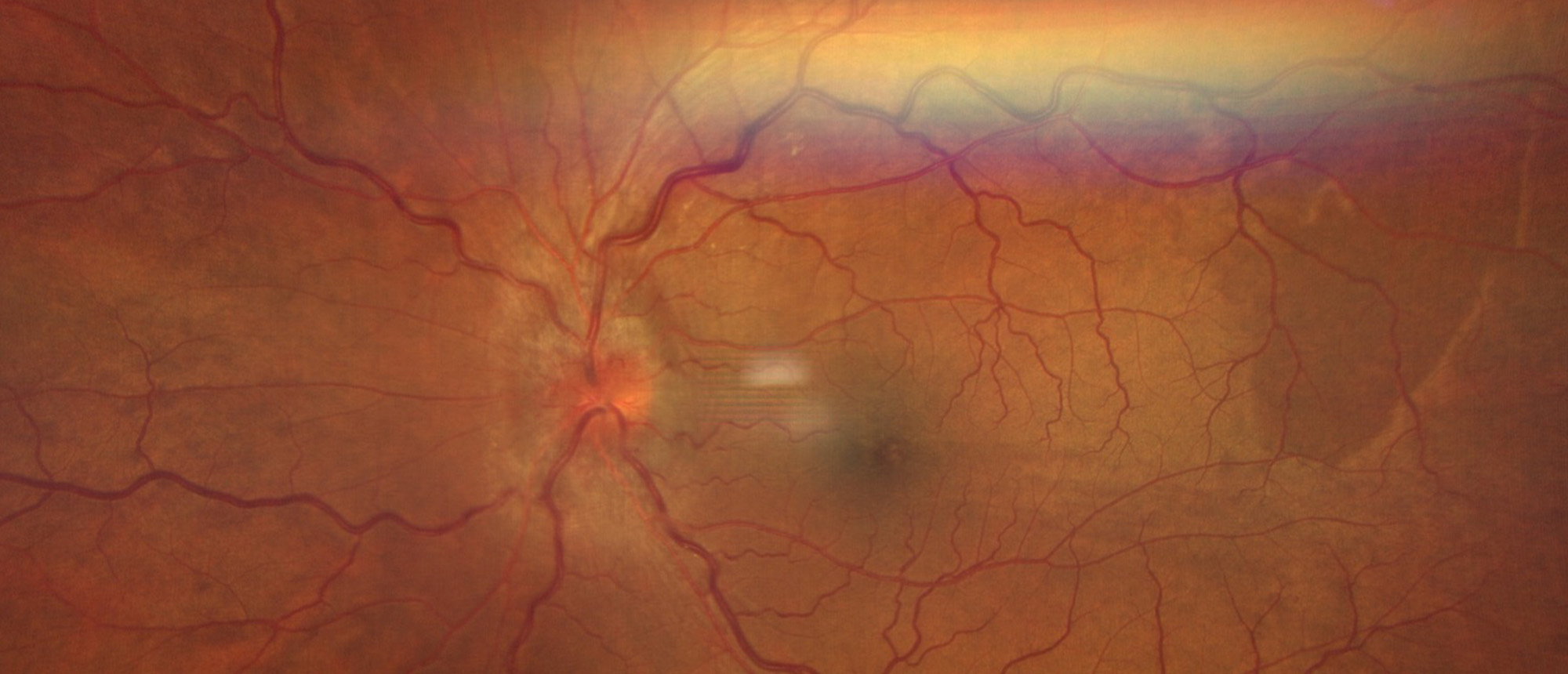
Thyroid Associated Ophthalmopathy with Compressive Optic Neuropathy
A patient presented for a second opinion after experiencing pain, proptosis, vision loss, and injection of his left eye for 2 years, with intermittent improvement on systemic steroids. Prior to presentation at our institution, the patient had MRI imaging, a cerebral angiogram, lab tests including normal ACE, TSH, T4, IgG subsets, ANCA, and quantiferon, and an unremarkable orbital biopsy. Upon presentation to our neuro-ophthalmology clinic, he was found to have proptosis, injection, chemosis, restricted extraocular movements, an afferent pupillary defect, and reduced visual fields. Imaging revealed enlargement of the extraocular muscles with sparing of the tendonous insertions. Repeat thyroid studies revealed a decreased TSH with normal T4 and TSI. Given medical comorbidities, patient was unable to undergo surgery, and thus received 15 days of intravenous methylprednisolone. The patient is pending teprotumumab approval.Thyroid ophthalmopathy is a disease most frequently associated with hyperthyroidism. In a small proportion of patients (approximately 5-6%), thyroid ophthalmopathy can be seen in euthyroid patients, making diagnosis challenging. A high index of suspicion should be maintained, and thyroid studies repeated. Findings on imaging that can lead to the diagnosis areenlargement of the muscle bellies with sparing of tendinous insertion. Patients with active disease benefit from steroid therapy, radiation therapy, teprotumumab therapy, or surgical intervention depending on the severity.
Presentation Date: 10/06/2022
Issue Date: 11/11/2022
Please log in or click on ENROLL ME to access this course.

