Polypoidal Choroidal Vasculopathy
Completion requirements
Description
Description
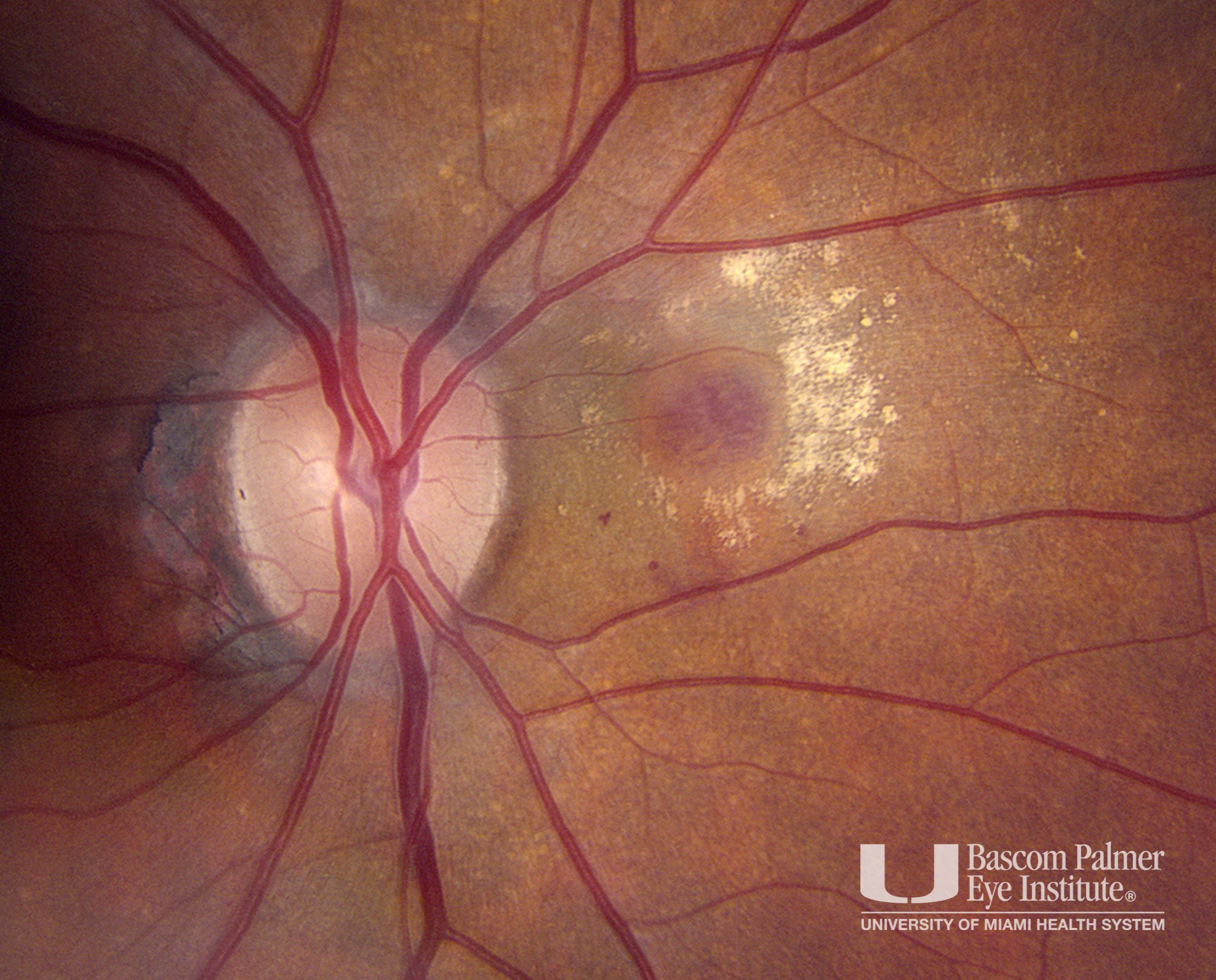
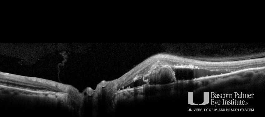
Fundus photo with a nasal peripapillary sub retinal vascular complex surrounded by exudation. OCT with pigment epithelial detachment and associated sub-retinal fluid. ICG angiography makes the diagnosis clear with a hypercyanescent polyp in the choroid.
Uploaded on: 07/16/2020