Choroideremia Associated with Choroidal Neovascularization
Completion requirements
Description
Description
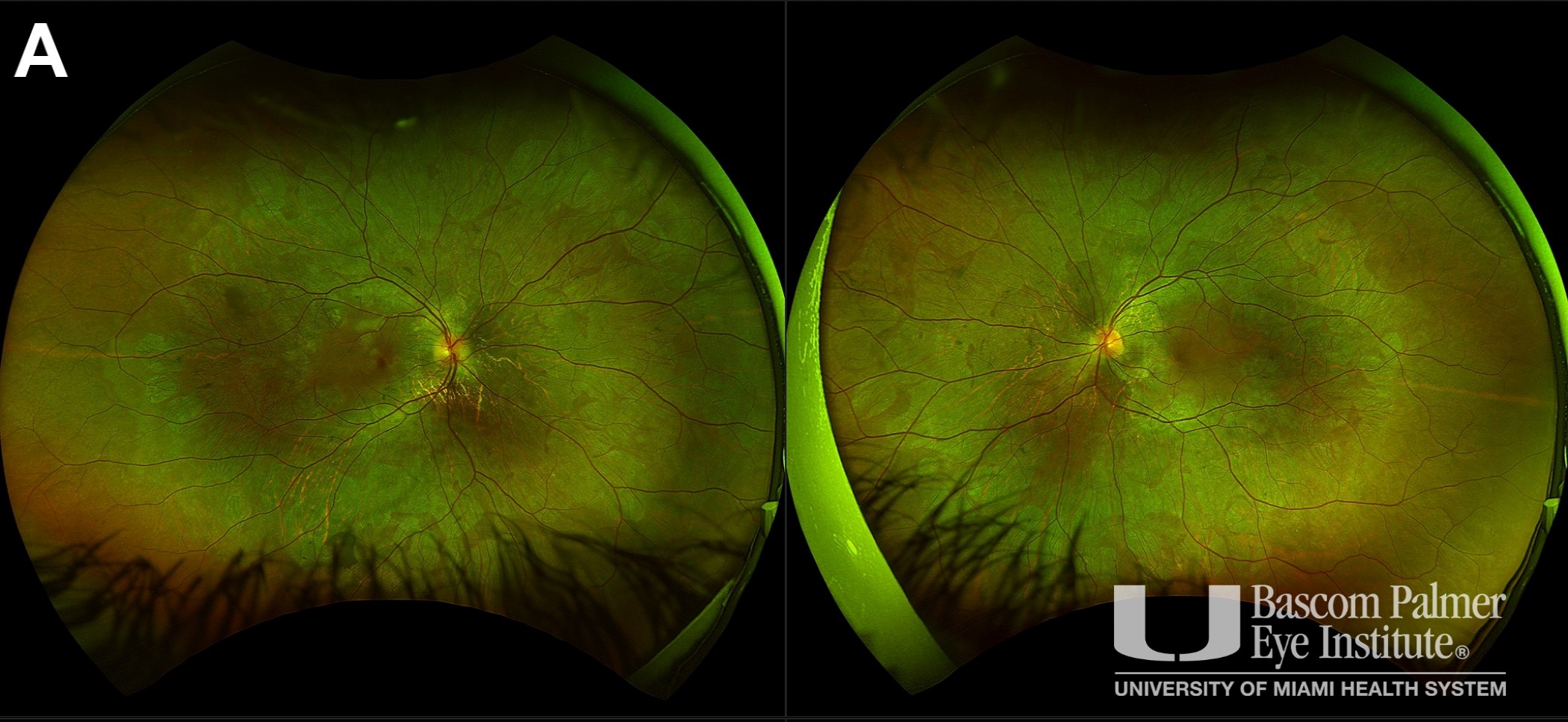
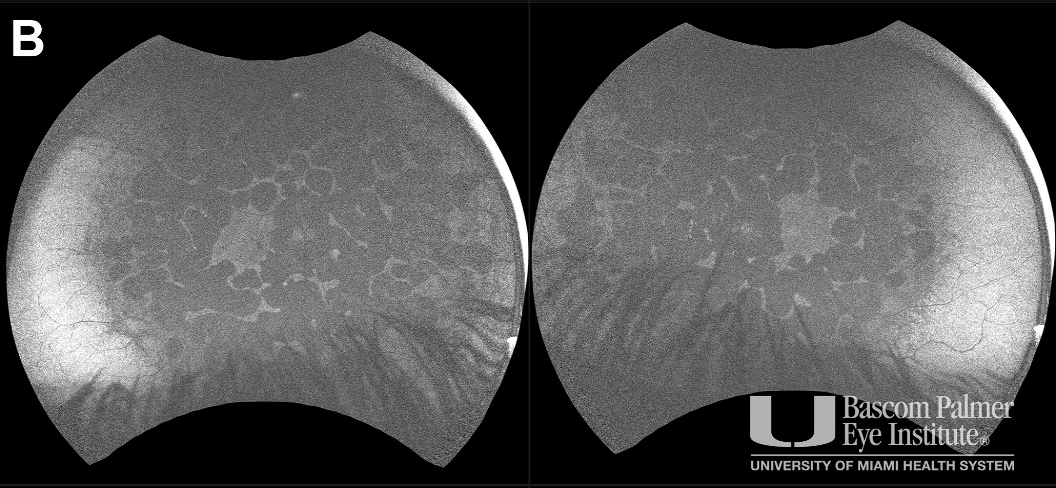
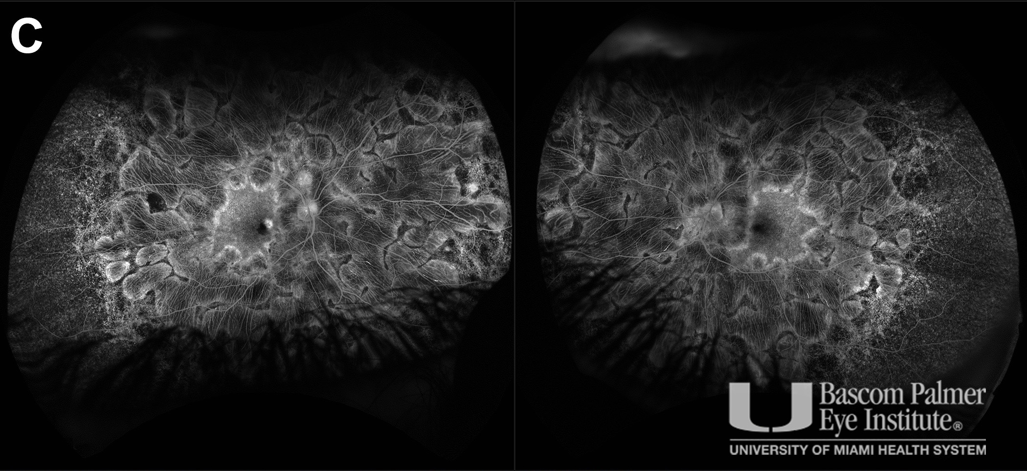
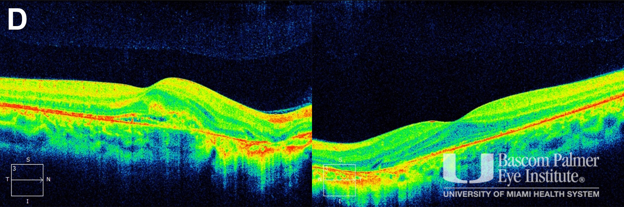
A patient presented with a central scotoma in the right eye. Fundus examination revealed midperipheral chorioretinal atrophy in both eyes (Fig A). Fundus autofluorescence demonstrated areas of confluent midperipheral hypoautofluorescence (Fig B). Optical coherence tomography demonstrated choroidal neovascularization with subretinal fluid in the right eye and parafoveal outer retinal atrophy in both eyes (Fig C). Fluorescein angiography demonstrated midperipheral window defects in both eyes and leakage in the right macula (Fig D). Genetic testing revealed a hemizygous exon 2 deletion on the CHM gene consistent with choroideremia.
Uploaded on: 03/27/2023