Bascom Palmer
  • Calendar
  • SPECIALTIES
    Cataracts and Intraocular Lenses Comprehensive Ophthalmology Corneal and External Diseases Glaucoma Neuro-Ophthalmology Ocular Oncology Ophthalmic Pathology Ophthalmic Plastic and Reconstructive Surgery Pediatric Ophthalmology and Strabismus Retina and Vitreous Diseases Uveitis
  • FILTER BY
    Online Grand Rounds Medical Student Education Ophthalmic Images Other Lectures Surgical Videos
  • SEARCH
  • More
Site-wide search Close
Toggle search input
guest access
Log in
Bascom Palmer
Calendar SPECIALTIES Collapse Expand
Cataracts and Intraocular Lenses Comprehensive Ophthalmology Corneal and External Diseases Glaucoma Neuro-Ophthalmology Ocular Oncology Ophthalmic Pathology Ophthalmic Plastic and Reconstructive Surgery Pediatric Ophthalmology and Strabismus Retina and Vitreous Diseases Uveitis
FILTER BY Collapse Expand
Online Grand Rounds Medical Student Education Ophthalmic Images Other Lectures Surgical Videos
SEARCH
Expand all Collapse all
  1. Dashboard
  2. CC092821OI
  3. Ophthalmic Image(s)
  4. Canthaxanthin Crystalline Retinopathy

Canthaxanthin Crystalline Retinopathy

Completion requirements

Description

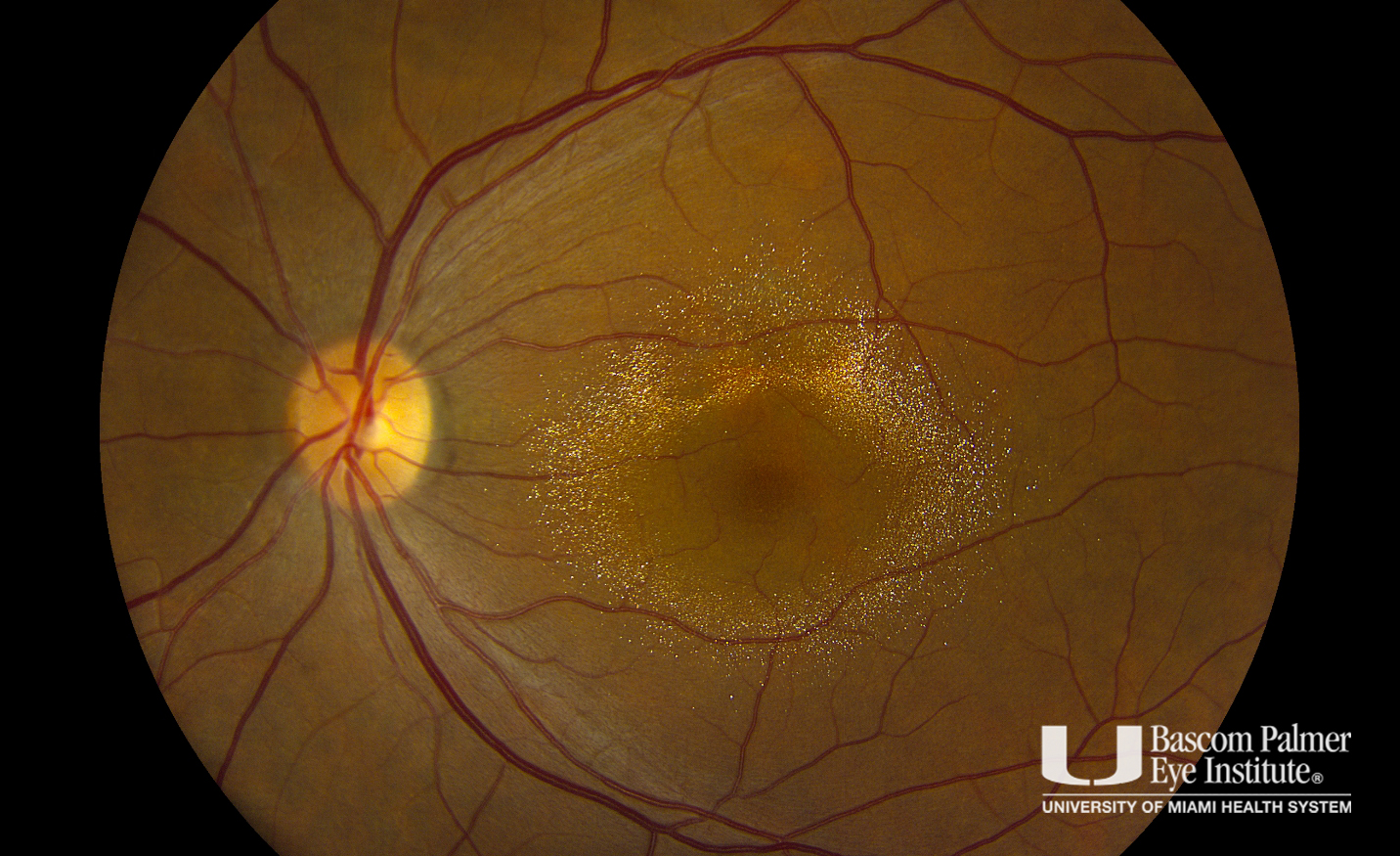
A patient with a history of progressive blurry vision in her left eye for 2 years prior to her presentation. She has no significant past medical except for consuming an over the counter oral tanning pills for a period of 7 years that was stopped 2 years prior to her presentation. Her exam was notable for a visual acuity of 20/20 in her right eye and 20/30 in the left eye. Anterior segment exam shows no abnormalities. Posterior segment exam shows scant intraretinal crystals in her right eye and a striking brilliant golden-yellow crystal deposition in a donut-shaped configuration around the center of the macula in her left eye. SD-OCT shows the deposition of crystals involving all retinal layers. Four years after her initial presentation, the patient continues to refrain from using the oral tanning agent and her visual acuity recovers back to 20/20 in the left eye. Posterior segment exam along with fundus and SD-OCT imaging shows partial resolution of the crystals more so in the left eye than the right, leaving a small area of PED and intraretinal cystic changes as evident in the SD-OCT image in her left eye which may signify a permanent damage from the crystalline retinopathy.

Uploaded on: 09/28/2021


Canthaxanthin Crystalline Retinopathy


◄ Alaa Al-Dabbagh, MD
guest access (Log in)
Data retention summary
Powered by Moodle
 
  • Medical Disclaimer
  • |
  • Terms of Use
  • |
  • Privacy Statement
  • |
  • HIPAA Notice of Privacy Practices
  • |
  • About Us
  • |
  • FAQs

© 2026 University of Miami Health Systems. All Rights Reserved.