MOGAD Optic Neuritis with Atypical Presentation of Roth Spots
Completion requirements
Description
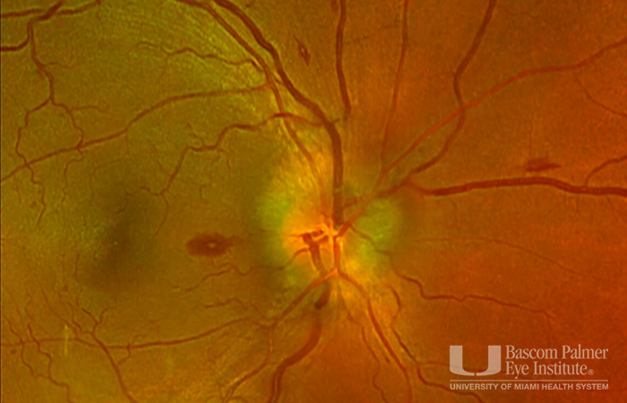
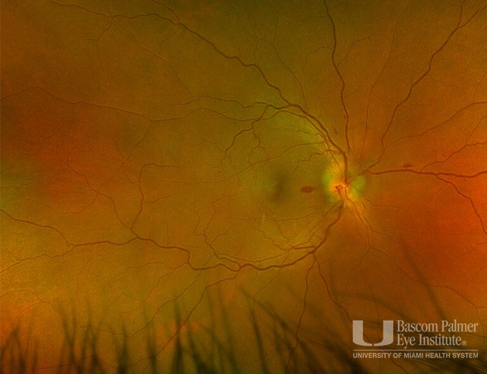
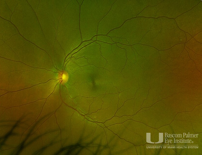
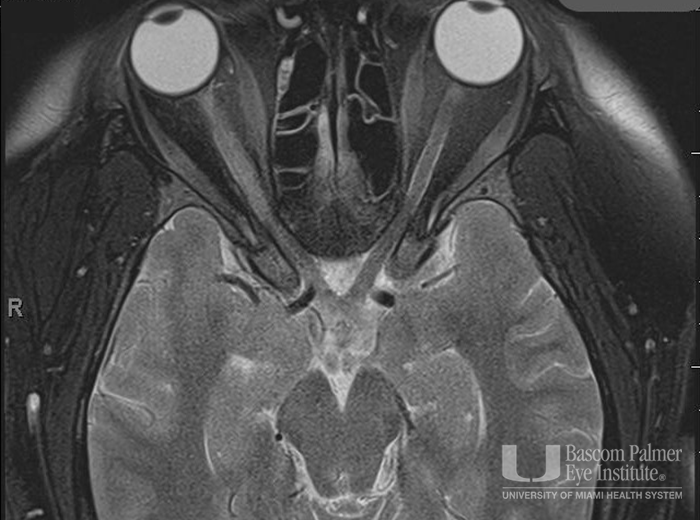
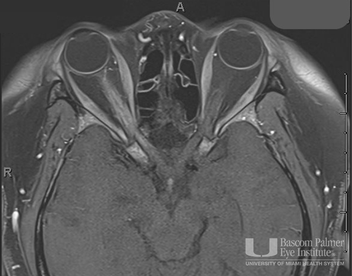
A patient with unilateral severe optic neuritis and ipsilateral retinal Roth spots. Fundus photos with significant optic nerve swelling in the right eye and multiple Roth spots in the same eye. Optic nerve head OCT of the right eye with Elevated disc contour, Expanded prelaminar tissue, Thickened neuroretinal rim appearance and Peripapillary subretinal fluid. Orbit MRI with Long segment swelling / enlargement of the right optic nerve with high T2 signal intensity and enhancement
Uploaded on: 04/30/2026