X-linked Retinoschisis
Section outline
-
-
Description
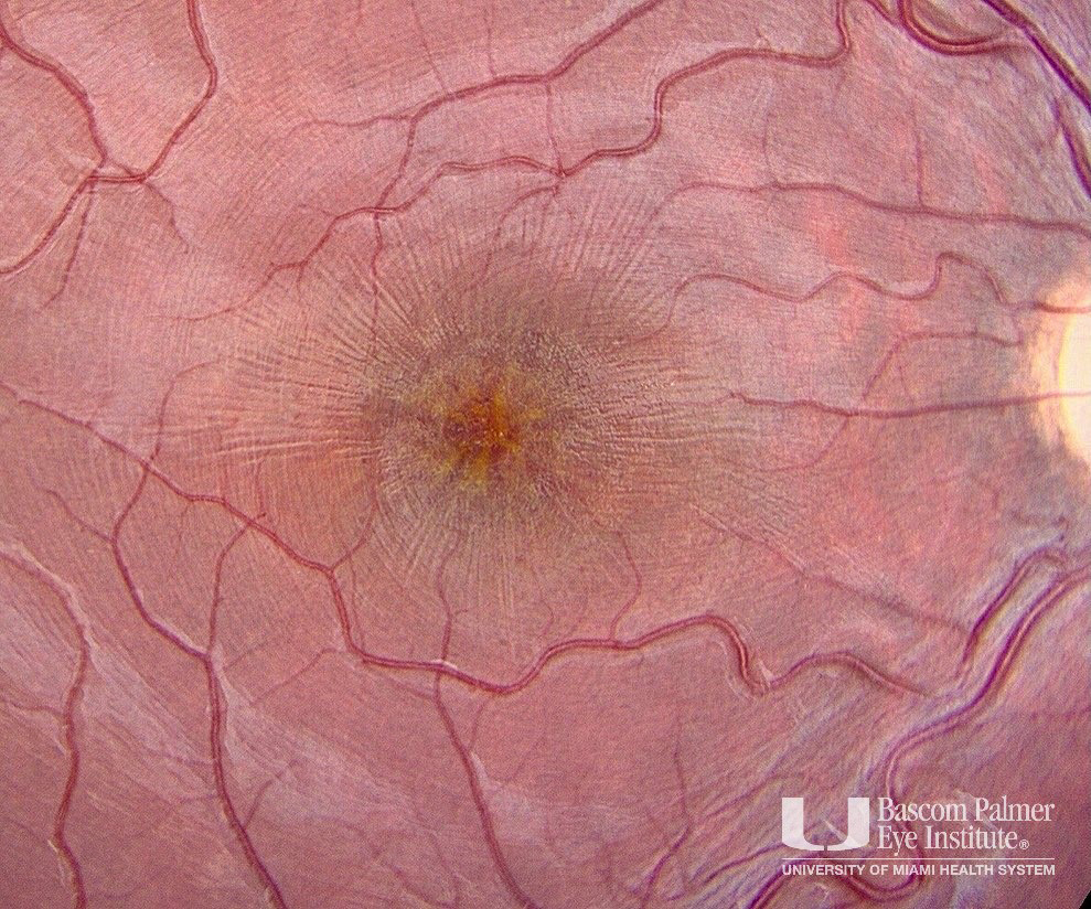
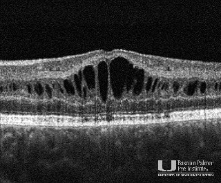
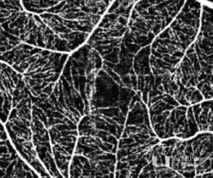
This is congenital retinal dystrophy is caused by a mutation in the retinoschisin (RS1) gene, which encodes for a protein that functions in cellular adhesion. The fundus photo shows a macular stellate pattern caused by schisis or splitting of the retinal layers at the level of the outer plexiform layer and outer nuclear layer. OCT and OCTA imaging reveal alterations of the foveal avascular zone in the superficial plexus, distortion of vessel architecture, and avascular cystic cavities. There are no telangiectasis vessels, nor aneurysmal dilations noted.
Uploaded on: 02/02/2021


