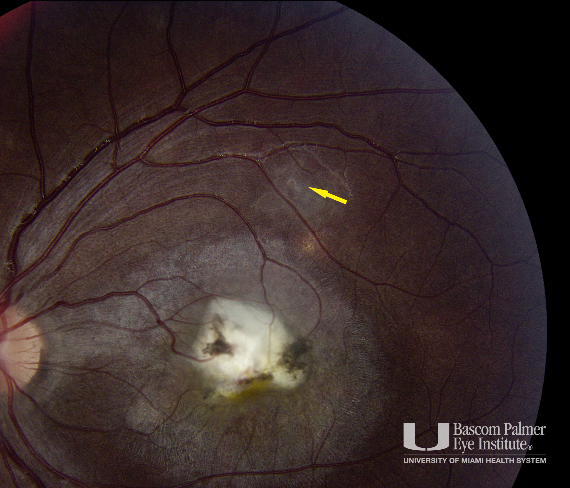
Diffuse Unilateral Subacute Neuroretinitis (DUSN) Treated With Laser
Section outline
-
-
Description
Fundus photo with multifocal unilateral white dots and central atrophy. On closer inspection a sub retinal nematode is found (yellow arrow). Post laser treatment with laser photocoagulation and with many years of follow up there was no progression of the uveitis.
Uploaded on: 07/16/2020

