Ophthalmic Image(s)
Section outline
-
-
Description
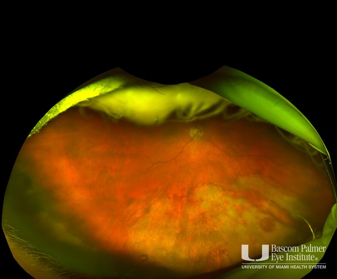
Right eye of patient who presented to clinic due to floaters, on fundus exam he had this peripheral whitish confluent granular lesion with areas of atrophy.
Uploaded on: 04/13/2021