Ophthalmic Image(s)
Section outline
-
-
Description
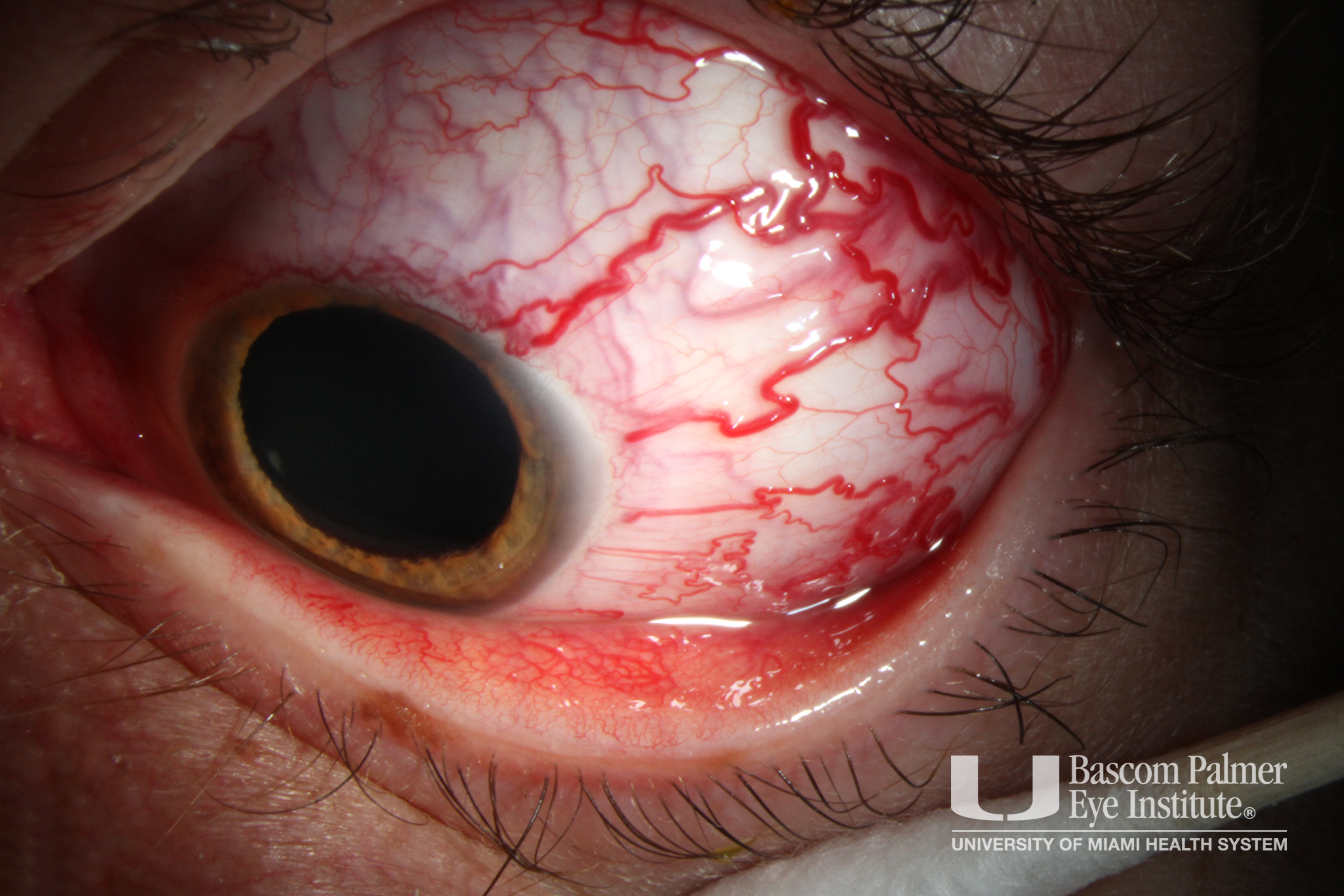
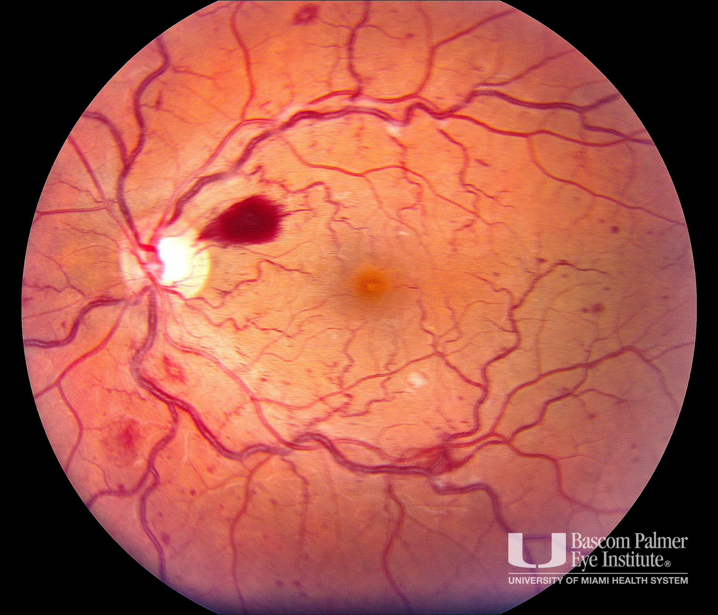
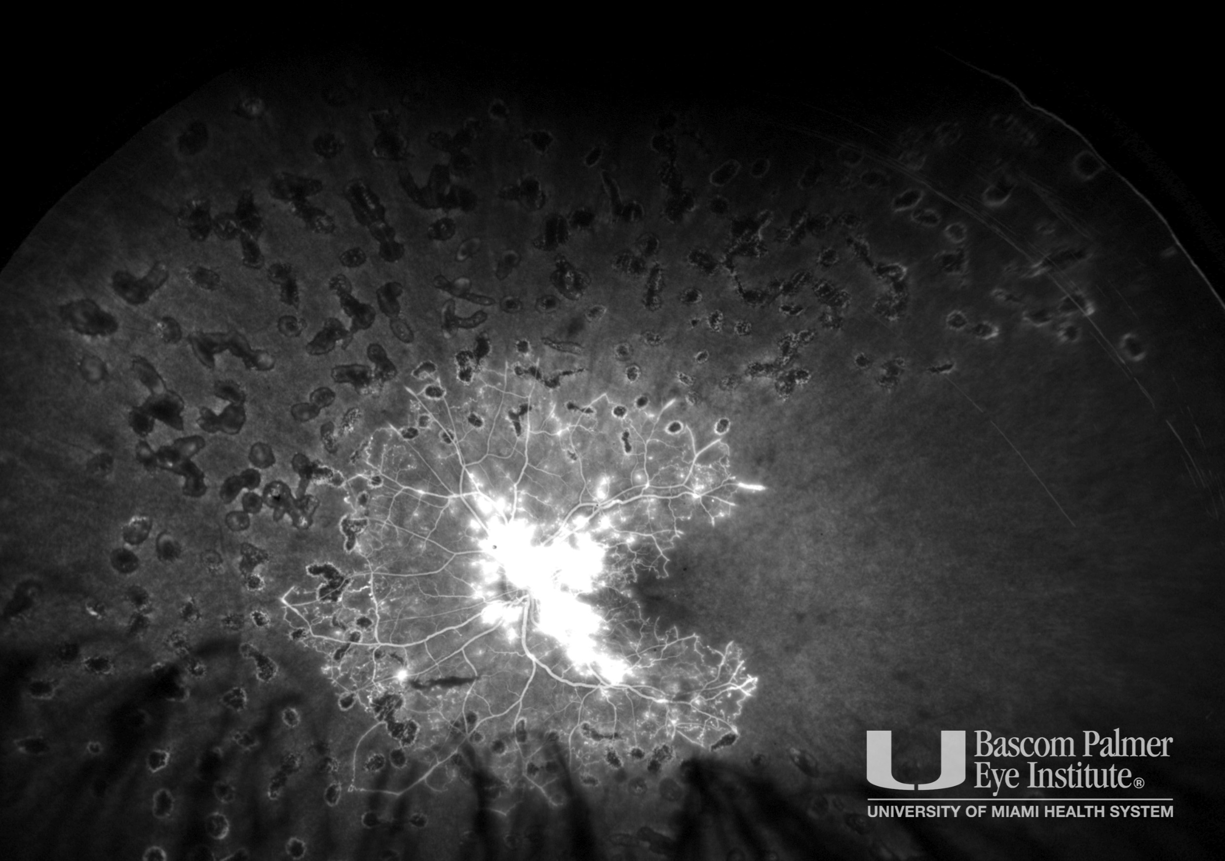
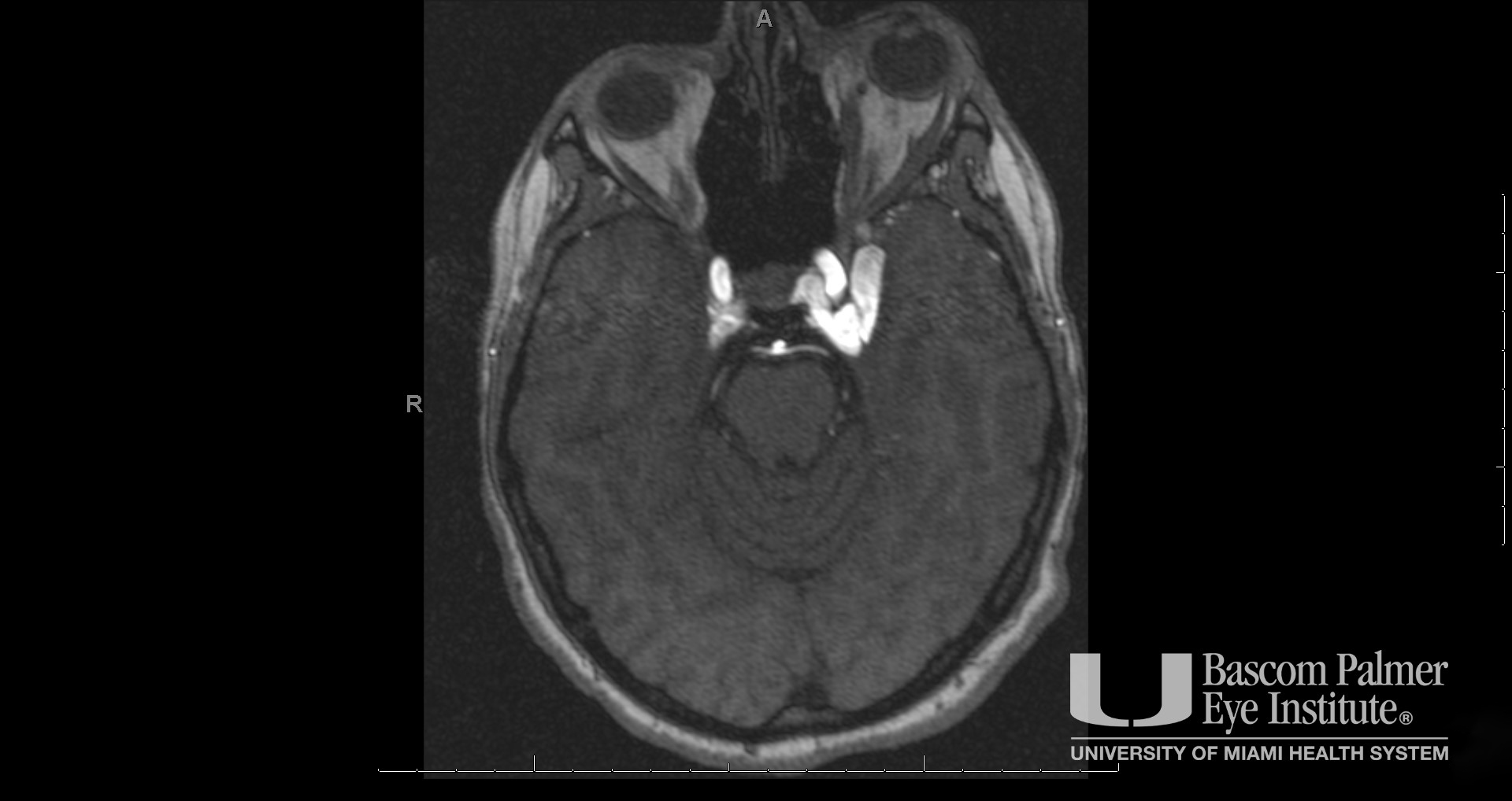
Slit lamp photo with dilated epi-scleral veins. Fundus photo with retinal hemorrhages. FA with severe non perfusion, leakage from the disc, and laser scars. MRI/MRA demonstrates left distal precavernous/proximal cavernous ICA fistula into the cavernous sinus with arterialized flow. There is resultant asymmetric enlargement of the left extraocular muscles and left globe proptosis.
Uploaded on: 07/16/2020